


INTRODUCTION
Liver cancer is the fifth most common cancer in men (7.9% of all cancers), theseventh most common cancer in women (6.5% of all cancers) [1], and the third most common cause of cancer death worldwide [2]. In Korea, liver cancer is the second leading cause of malignancy mortality, and ~22.5 per 100,000 individuals died of hepatocellular carcinoma (HCC) [3]. Despite recent progress in follow-ups and treatment strategies, the overall prognosis of patients with HCC has remained dismal (5-year survival rate, <10%) owing to the high proportion of patients with advanced HCC [3].
If HCC is diagnosed at its late or advanced stage, only palliative treatment, such as trans-arterial chemoembolization, molecular targeted agent administration, or immunotherapy [4] can be applied, which results in poor survival. However, if HCC is recognized at its early stage, curative treatment options, such as resection, percutaneous ablation, or orthotopic liver transplantation, can be considered, which might yield favorable long-term outcomes [5-8]. For these reasons, various HCC prediction models, such as the Chinese university (CU)-HCC and risk estimation for HCC in chronic hepatitis B (CHB) (REACH-B) models, have been proposed for early HCC detection in patients with CHB [2]. Recently, magnetic resonance imaging (MRI) using the new liver-specific contrast agent gadoxetic acid (Primovist) have been widely used for HCC diagnosis [9,10].
Recently, Cho et al. [11] proposed a risk prediction model for the development of HCC from indeterminate nodules detected on computed tomography (CT) (RadCT score) in patients with CHB-related cirrhosis. Unlike the CU-HCC or REACH-B model, this risk model includes radiological findings of “arterial enhancement” on CT [11]. In their study, indeterminate nodules were categorized into three risk groups with significantly different HCC risks (low-risk group, 1%; intermediate-risk group, 14.5%; and high-risk group, 63.1% at 5 years). The Liver Imaging Reporting and Data System (LI-RADS) [12] subdivides hepatic nodules detected on CT and MRI in patients at a high risk for HCC based on their radiological characteristics; indeterminate nodules are subdivided as “probably benign (LR 2),” “intermediate probability (LR 3),” and “probably HCC (LR 4)” [12]. In a previous study, patients with CHB-related liver cirrhosis and indeterminate nodules showed significantly higher risks of HCC development [11].
PATIENTS AND METHODS
Patients
Patients with CHB who had LI-RADS 2/3 lesions on MRI conducted at Yonsei Liver Center, Severance Hospital, Yonsei University College of Medicine from 2013 to 2016 were eligible for inclusion in this retrospective cohort study. CHB was defined as chronic necroinflammatory disease of the liver caused by persistent infection with hepatitis B virus (HBV), with presence of HBV surface antigen in the serum for at least 6 months. LI-RADS 2/3 nodules were defined on the basis of LI-RADS nodules detected on MRI performed for both primary HCC surveillance and diagnosis to characterize liver lesions identified on ultrasonography during HCC surveillance [12-15].
The exclusion criteria were as follows: (1) HCC development <6 months after enrollment; (2) indeterminate nodules related to previously treated lesion; (3) co-infection with hepatitis C and human immunodeficiency virus; (4) active extrahepatic malignancies; (5) other significant medical illness; (6) significant alcohol consumption (>40 g/daily); and (7) insufficient clinical or radiological information for statistical analysis.
The study conformed to the ethical guidelines of the 1975 Declaration of Helsinki and was approved by the institutional review board of each institute. The requirement for written informed consent was waived owing to the retrospective nature of the study.
Baseline data collection
Information on age; sex; other co-morbidities, such as hypertension or diabetes mellitus; body mass index; liver cirrhosis ongoing antiviral therapy (AVT); and HCC history, was collected from the medical records. Baseline laboratory parameters, including levels of HBV envelope antigen (HBeAg), HBV DNA, alpha-feto-protein (AFP), aspartate aminotransferase (AST), alanine aminotransferase (ALT), total bilirubin, and serum albumin and platelet counts, were collected. Radiological findings of nodules, such as maximal size, arterial enhancement, T2 hyperintensity, and diffusion restriction, were also reviewed. The imaging analysis results were interpreted by two expert abdominal radiologists. If the results were different, the final decision was made after agreement between them.
RadCT score
The risk model for the development of HCC from indeterminate nodules was recently proposed by Cho et al. (RadCT score).11 This score is composed of seven factors: age, enhancement pattern, size, serum albumin level, serum AFP level, HCC history, and HBeAg level. The formula for the risk score calculation was as follows.
Risk score=1×age (years)+19×enhancement pattern (arterial non-enhancement or arterioportal shunt=0; arterial enhancement =1)+42×size (≤1 cm=0; >1 cm=1)+16×serum albumin level (>3.5 g/dL=0; ≤3.5 g/dL=1)+31×serum AFP level (<100 ng/mL=0; ≥100 ng/mL=1)+32×HCC history (no=0; yes=1)+18×HBeAg level (negative=0; positive=1)
Two cutoff values (60 and 105) discriminated the HCC risk into three categories. The 3-year HCC development rate was 1%, 14.5%, and 53.3%, and the 5-year HCC development rate was 1%, 14.5%, and 63.1% in the low-, intermediate-, and high-risk groups, respectively.
CU-HCC and REACH-B scores
The CU-HCC score was first derived in a cohort of 1,005 Chinese patients with CHB [16,17]. It is composed of five factors: age, serum albumin level, total bilirubin level, HBV DNA level, and cirrhosis and ranges from 0 to 44.5. Two cutoff values (5 and 20) discriminated the HCC risk into three categories. The 5-year HCCfree survival rates were 98.3%, 90.5%, and 78.9% in the low-, medium-, and high-risk groups, respectively.
The REACH-B score was derived in 3,584 Chinese patients with CHB from the Taiwanese Risk Evaluation of Viral Load Elevation and Associated Liver (REVEAL) cohort and validated in a cohort of 1,505 patients from three tertiary referral clinics in Hong Kong and South Korea [16,18]. The variables included in the risk score are sex, age, serum level of ALT, level of HBeAg, and level of HBV DNA. A 17-point risk score was developed, and the HCC risk rate ranged from 0% to 47.4% at 5 years and from 0% to 81.6% at 10 years for patients with the lowest and highest HCC risks, respectively.
Follow-up and HCC diagnosis
Although patients with HCC history were included, all participants were confirmed as being HCC-free at the beginning of the study. Each patient was followed up with AFP examination and ultrasonography, CT, or MRI every 3 or 6 months for HCC surveillance. Most of the patients have been followed up with ultrasonography every 6 months. However, the high-risk patients, for instance, who showed equivocal results based on ultrasonographic findings and AFP level received dynamic imaging according to the physicians’ decision. Protocols of CT and MRI are described in Supplementary Material. During the follow-up, data on the actual development to HCC were collected. Two radiologists reviewed the dynamic CT and MRI imaging. HCC diagnosis was based on the radiological findings on CT and/or MRI or on the histological evidence according to the guideline of the American Association for the Study of Liver Diseases [19].
Statistical analysis
Student’s t-test (or Mann-Whitney test) and the chi-squared test (or Fisher’s exact test) were used to compare the baseline characteristics of the patients. In this analysis, we checked the association of HCC occurrence with each variable. The patients were censored at the time of first presentation of HCC or at the last follow-up. The annual and cumulative incidence of HCC was calculated using the Kaplan-Meier method. To identify the independent risk factors for HCC development, univariate and subsequent multivariate Cox proportional hazard regression analyses were conducted. Hazard ratios (HRs) and corresponding 95% confidence intervals (CIs) were presented. The final risk score obtained in the RadCT and conventional prediction models, including the CU-HCC and REACH-B models, was validated in this cohort. Each risk model was validated via Cox regression analyses. All statistical analysis was performed using the IBM SPSS statistics software, version 24 for Windows (IBM Corp., Armonk, NY, USA).
RESULTS
Baseline characteristics
Among 2,748 consecutive patients who underwent liver MRI during the study period, there were 180 patients with LI-RADS 2/3 lesions. After excluding 81 patients, 99 patients were selected for the final statistical analysis (Fig. 1).
The baseline patient characteristics are described in Table 1. The median age was 58 years, and the male sex predominated (n=72, 72.7%). Liver cirrhosis and HCC history were identified in 44 (44.4%) and 47 (47.5%) patients, respectively. Of the 47 patients who had a previous HCC history, 28 patients received curative treatment (operation [n=22], radiofrequency ablation [n=5], and cryoablation [n=1]) and the other 19 patients received noncurative treatment (trans-arterial chemoembolization) with the achievement of complete response. The median maximal tumor size was 11.0 mm. Arterial enhancement, T2 hyperintensity, and diffusion restriction were identified in 16 (16.2%), 24 (24.2%), and 23 (23.2%) patients, respectively. The median CU-HCC, REACH-B, and RadCT scores were 6 (interquartile range [IQR]=3- 18), 8 (IQR=7-10), and 112 (IQR=90-130), respectively.
Comparison between the patients with and without HCC
During the follow-up period (median, 20 [IQR=8-29] months), the liver lesions in 41 (41.4%) patients progressed to HCC. All HCC diagnosis was made according to radiological findings. The comparison of the baseline characteristics between the patients with and without HCC is shown in Table 2. The disease-free survival of patients with HCC was significantly shorter than those without (median 30 vs. 46 month, P<0.001), but the overall survival was not significantly different (median 61 vs. 59 months, P=0.791).
The patients with HCC had significantly higher proportion of liver cirrhosis (68.3% vs. 58.6%) and HCC history (65.9% vs. 34.5%) (both P<0.05) than those without. They also had significantly lower AST (median, 29 vs. 36 IU/L) and ALT levels (median, 26 vs. 29 IU/L) and higher RadCT score (median, 120 vs. 98) than those without (all P<0.05). The CU-HCC and REACH-B scores were significantly similar between them (all P>0.05).
Unadjusted HR of the prediction models for HCC development
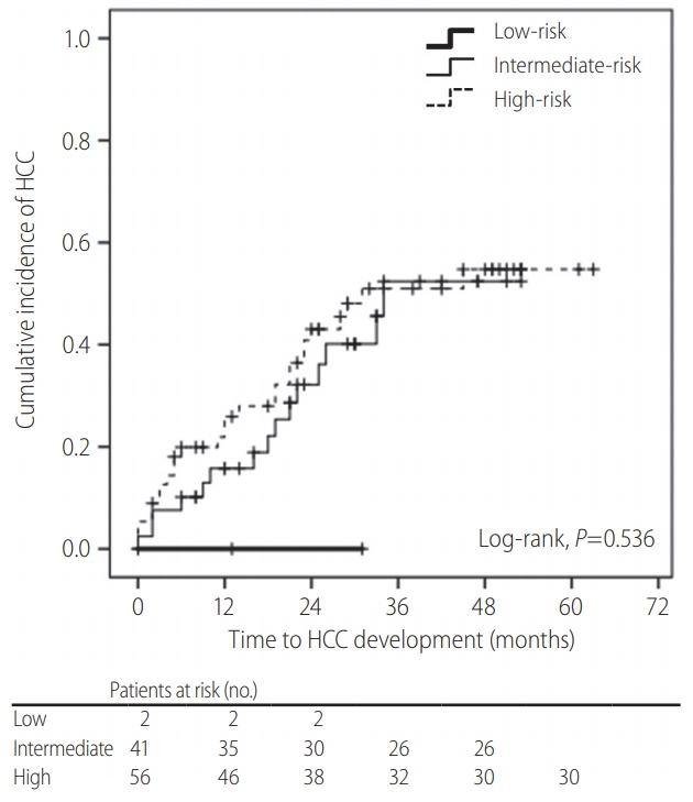
In contrast to those of the CU-HCC and REACH-B scores (both P>0.05), the unadjusted HR of the RadCT score was statistically significant for HCC development (HR=1.018, 95% CI=1.005- 1.031, P=0.007) (Table 3). However, when the subjects were stratified into three risk groups based on the RadCT score (<60, 60–105, and >105), the cumulative HCC incidence was not significantly different among them (overall P=0.536, all P>0.05 among adjacent curves, log-rank test) (Fig. 2).
Independent predictors of HCC development
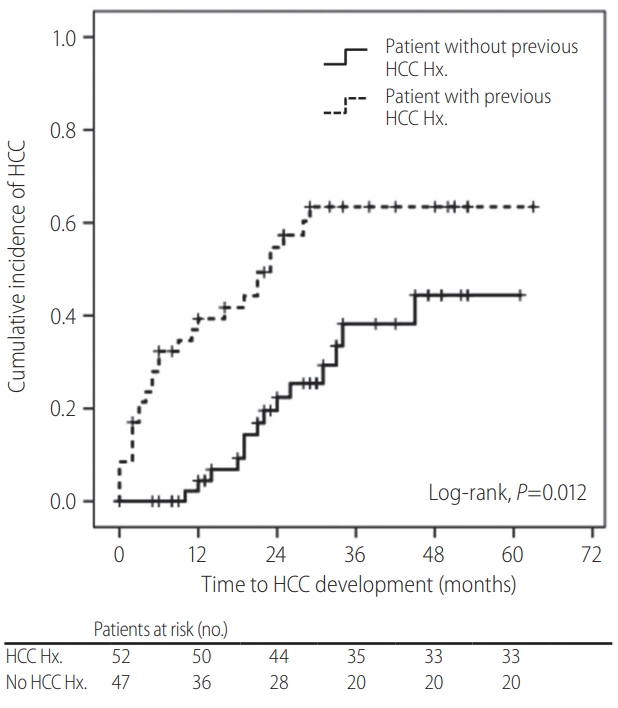
The Cox regression analysis performed to identify the risk factors associated with development from LI-RADS 2/3 nodules to HCC is shown in Table 4. The univariate analysis revealed that liver cirrhosis, HCC history, and the RadCT score were associated with HCC development risk (all P<0.05). In contrast, the MRI parameters, including nodule size, arterial enhancement, T2 hyperintensity, and diffusion restriction, were not predictive of HCC development in the univariate analysis (all P >0.05). In the subsequent multivariate analysis, HCC history was the only independent predictor of HCC development (HR=2.374; 95% CI=1.205-4.678; P=0.012). When the subjects were divided into two groups according to HCC history, the cumulative incidence of HCC was significantly higher in the patients with HCC history than in those without (4.2% vs. 23.6% at 1 year; 33.5% vs. 63.4% at 3 years; P<0.001, log-rank test) (Fig. 3).
DISCUSSION
An accurate assessment of the risk of HCC development is important for establishing individualized strategies of follow-up, intervention, and management because this ultimately enables the extension of overall survival in patients with CHB [2]. To address this issue, several risk prediction models for patients with CHB have been proposed [2,17,20]. In addition, the importance of dysplastic nodules as precancerous lesions of HCC is well established. Sakamoto et al. found that multistep carcinogenesis is one pathway to HCC development based on 320 resected liver tissues [21]; other investigators also suggested that macroregenerative nodules may represent precancerous lesions [22,23]. However, because hepatic nodular lesions do not necessarily progress to HCC, [24-26] it is important to identify true precancerous lesions. Thus, a new risk prediction model (RadCT score) for the development of HCC from indeterminate nodules on CT in patients with CHB-related cirrhosis has been proposed [11].
In this study, we attempted to validate the prognostic accuracy of the RadCT score. Although the unadjusted HR of the RadCT score was statistically significant (HR=1.018, P=0.007), the cumulative risk of HCC development was not significantly different among the risk groups after adjustment. Instead, HCC history, one of the constituent variables of the RadCT score, was the only predictor of HCC development. The CU-HCC and REACH-B scores did not significantly predict the risk of HCC development. This finding suggests that the RadCT score, a new CT-based risk prediction model, and conventional risk models for HBV-related HCC development in patients with CHB-related cirrhosis, might not be applicable for LI-RADS 2/3 nodules on MRI. Thus, further studies are required to optimize the cutoff value for each risk group or establish another MRI-based prediction model.
Our study has several limitations and strengths. First, we validated the predictive performance of the RadCT score for indeterminate nodules in patients with CHB-related cirrhosis and investigated the prognostic accuracy of other conventional risk prediction models. Kobayashi et al. [27] reported that 18.8% of 154 nodules in patients with chronic liver disease progressed to HCC during their follow-up period; the cumulative rate of HCC development at 5 years was 80.0% for high-grade; 36.6%, low-grade; and 12.4%, regenerative dysplastic nodules. However, because histological assessments for indeterminate liver nodules are generally infeasible, [28] imaging-based risk assessments, such as use of the RadCT score, which is composed of routinely available clinical variables, might be appropriate in clinical practice; however, its predictive accuracy was not validated in our study.
Second, we focused on indeterminate nodules without a definite diagnosis of HCC, which were classified as “probably benign (LR 2)” and “intermediate probability (LR 3)” according to the LIRADS guidelines. LR 2/3 nodules can progress to HCC [27]. However, in contrast to LR 1 nodules without malignancy risks and LR 4/5 nodules with high HCC risks, the probability of HCC development is in the gray zone, with a relatively low risk of HCC development. In our study, 76 out of the 99 patients had only one LI-RADS 2/3 lesion, whereas the others have more than two LI-RADS 2/3 lesions (two in 16 and three in 7 patients). Although several LIRADS 2/3 lesions can be found simultaneously in a given patient, we considered HCC development as the solid end-points in our study, regardless of the number of LI-RADS 2/3 lesions. Indeed, the aim of our study was to identify the risk factors of HCC development among patients with LI-RADS 2/3 lesions in a clinical point of view, not to identify the risk factors of HCC development among LI-RADS 2/3 lesions in a radiological point of view. Thus, it is important to investigate whether the risk prediction models for LR 2/3 nodules can identify high-risk patients who would benefit from more careful monitoring, which might support the rationale of our study.
Third, although the predictive accuracy was not satisfactory, we tested whether the well-known CU-HCC and REACH-B models have similar prognostic performances when indeterminate nodules exist in patients with CHB. However, these two models were not associated with the risk of HCC development even in the univariate analysis in our cohort; this indicates that the CU-HCC and REACH-B models might not be applicable in this clinical setting. This finding can be explained in several ways. First, despite the low risk of HCC development on MRI, the presence of liver nodules might indicate that the patients already have a higher risk of HCC development than those in the cohorts from which the CUHCC and REACH-B models were derived. Second, because we attempted to validate the RadCT score, we included patients with HCC history to maintain similar clinical characteristics of the study cohort. Although we excluded patients who developed HCC immediately after enrollment, HCC history is a strong risk factor of HCC development [11,29]. Indeed, HCC history was the only predictor of HCC development in our study.
There were also several limitations in our study, which remained unresolved. First, the CU-HCC and REACH-B models were basically generated from the cohorts with CHB patients without radiological information regarding the presence of indeterminate hepatic nodules. This might indicate that the CU-HCC and the REACH-B models might have acceptable predictive accuracy, regardless of the potential presence of indeterminate hepatic nodules. Our study focused on only CHB patients with indeterminate hepatic nodules by dynamic imaging, who might be not recognized as having indeterminate hepatic nodules by ultrasound. Thus, our study population might be a subgroup of the whole CHB patients. In addition, due to the different clinical characteristics from the cohorts where CU-HCC and REACH-B models were derived, it might be biased to use CU-HCC and REACH-B models as controls. However, as these two models are well-known predictive models, we choose them for comparison.
Second, previous HCC history was selected as a significant predictor of HCC development in our study. Because Khalili et al. [30] reported that around 20% among 1–2 cm-sized indeterminate nodules were finally found to be HCC, our results should be interpreted cautiously. Although we included patients who showed HCC-free status for at least 6 months to maximize small sample size, further large-scale studies are warranted to resolve this issue.
Third, despite the large cohort of patients with CHB before exclusion, only a small proportion was selected for the statistical analysis (n=99, 3.6%), and the follow-up period was relatively short (median, 20 [IQR=8–29] months). In addition, owing to the relatively small sample size, the constituent variables of the RadCT score, such as age, enhancement pattern, size, and serum albumin, serum AFP, and HBeAg levels, were not predictive of development from LI-RADS 2/3 nodules to HCC; this might explain why the RadCT score was significant only without adjustment.
Fourth, liver cirrhosis was not associated with the risk of HCC development in our study after adjustment (P=0.107). Although we do not know the exact reason, higher proportion of patients with AVT which can modify fibrotic burden in spite of morphological cirrhosis and small sample size of our cohort might explain this phenomenon.
In conclusion, our study validated the prognostic accuracy of a newly proposed risk prediction model for development of HCC from indeterminate nodules on CT (RadCT score); we found that the RadCT score was not predictive of HCC; conversely, HCC history predicted CHB-related development of HCC from LI-RADS 2/3 nodules. New risk models optimized for MRI-defined indeterminate nodules are required.

 PDF Links
 PubReader
 ePub Link
 Full text via DOI
 Full text via PMC
 Download Citation
 Supplement
 Print
Print


