| Clin Mol Hepatol > Volume 18(4); 2012 > Article |
Combined hepatocellular carcinoma and cholangiocarcinoma (combined HCC-CC) is a rare subtype of liver cancer displaying components of both hepatocellular and cholangiocellular carcinoma.1 Not only are exophytic liver tumors very rare, but their diagnosis presents a challenge due to the uncertainty of the tumor origin. In this issue, we present a case of exophytic combined HCC-CC with unusual morphologic features treated by liver transplantation. This study highlights the need for an increased awareness of the unusual morphologic features of this tumor to prevent potential misdiagnoses.
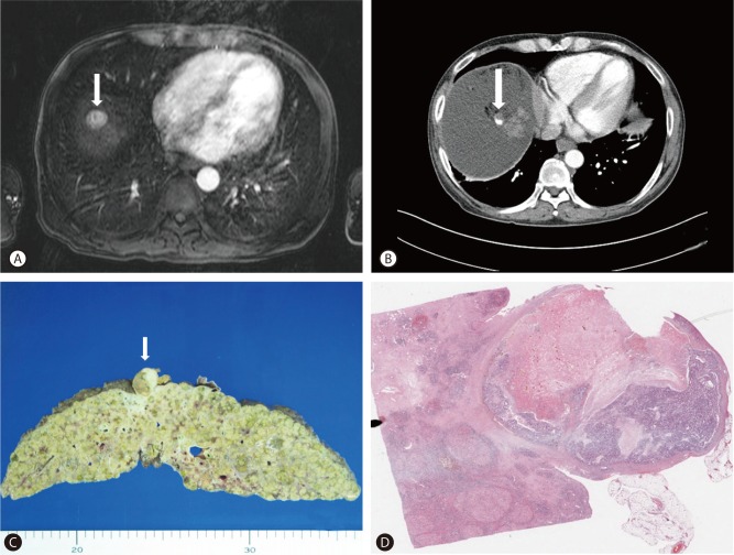
A 44-year-old man presented with a history of hepatitis B virus-related cirrhosis. A computed tomography (CT) scan revealed a 2 cm mass in segment 8 of the liver. The mass showed arterial hypervascularity and was washed out in the venous phase on enhanced CT. On the T2-weighted magnetic resonance (MR) image, a hyperintense focus was seen within a low-signal-intensity background nodule. On the gadolinium-enhanced MR image, this focus of HCC showed arterial enhancement and delayed washout (Fig. 1A). According to the American Association for the Study of Liver Disease criteria, the mass was diagnosed as HCC.2 Transarterial chemoembolization (TACE) using lipiodol mixed with adriamycin followed by the injection of gelatin sponge particles was performed. On follow-up CT images obtained 7 and 10 months after TACE, a non-lipiodolized portion in the anteroinferior aspect of the mass was detected and showed subtle enhancement on an arterial phase image with an increased size from 4├Ś2 mm to 13├Ś7 mm (Fig. 1B). The patient subsequently underwent liver transplantation (LT).
The transplanted liver tissue revealed a 2├Ś2 cm exophytic mass in the right liver (Fig. 1C, D). Histologically, the tumor consisted predominantly of tubular adenocarcinoma (~90%) with large areas of coagulation necrosis, and focal moderately or poorly differentiated HCC cells (~10%) arranged in a trabecular pattern. Between tumor cell nests, a sinusoidal pattern of blood vessels was noticed (Fig. 2A-D). Combined HCC-CC usually contained a variable number of tumor cells with intermediate morphology between HCC and CC within a desmoplastic stroma.1 However, there were a few tumor cells demonstrating morphology resembling an intermediate between HCC and CC, and desmoplastic reaction was minimal in the present tumor. Immunohistochemical staining showed that the adenocarcinoma tumor cells were positive for biliary markers keratin 7 and 19, and progenitor cell markers EpCAM, CD133, and CD56, whereas tumor cells with the trabecular pattern were positive for HepPar1 and EpCAM (Table 1 and Fig. 2E-H). Immunostaining for CD34 highlighted characteristic sinusoidal patterns of vascular structure, a typical blood vessel pattern of HCC, in the adenocarcinoma areas (Fig. 2I). According to the World Health Organization (WHO) definition and immunohistochemical findings, the tumor was diagnosed as transitional type combined HCC-CC. Twenty months after LT, the patient remained well, and a follow-up CT scan showed no recurrent cancer.
Combined HCC-CC is a rare subtype of liver cancer displaying components of both hepatocellular and cholangiocellular carcinoma.1 If the center of a tumor lies beyond the confines of the liver and the tumor originates from the liver, it can be defined as an exophytic hepatic tumor. Benign tumors such as hemangioma, hepatic adenoma, focal nodular hyperplasia, and angiomyolipoma and malignant tumors such as hepatocellular carcinoma, cholangiocellular carcinoma, and metastasis may show exophytic growth.3 Exophytic combined HCC-CC of the liver is unusual. The WHO classification defines the classical type of combined HCC-CC as a tumor containing unequivocal elements of both HCC and CC, which are intimately admixed. This tumor stands in a class of its own and should be distinguished from either HCC or CC arising in the same liver.1 Combined HCC-CC is divided into transition type and intermediate type.4 Both the transitional and intermediate type of combined HCC-CC contains a variable number of tumor cells with morphology resembling an intermediate between HCC and CC. Tumor cells with intermediate morphology consists of strands/trabeculae of small, uniform, oval-shaped cells with scant cytoplasm and hyperchromatic nuclei embedded within an abundant stroma, or proliferating tumor cells with an antler-like anastomosing pattern resembling the canal of Hering within a desmoplastic stroma, or solid nests of intermediate hepatocyte-like cells surrounded by small cells in the periphery.1,2,5
In our case, the combined HCC-CC exhibited unusual features as follows. First, the tumor was exophytic. Second, there were a few foci of tumor cells with intermediate morphology in a viable tumor component, which usually appeared in the combined HCC-CC. Third, an unusual sinusoidal pattern of tumor vessels and minimal desmoplastic stroma in cholangiocarcinoma areas were observed. Explanation for these unusual histologic features for this combined HCC-CC is unclear. We hypothesized that the combined HCC-CC phenotype of this tumor was obtained after the TACE treatment for HCC. This notion is supported by the fact that HCCs can acquire biliary phenotypes after TACE treatment.6,7 Zen et al demonstrated that HCC with the combined hepatocholangiocellular phenotype appears in post-TACE HCC.6 TACE could induce a more aggressive form of HCC characterized by a biliary phenotype and possibly derived from hepatic progenitor cells.7 Nishihara et al also suggested that the biliary phenotype of HCC originates from the adaptive transformation of the unaffected or TACE-resistant tumor cell population.8 Regarding the sinusoidal vessel pattern in adenocarcinoma areas of this tumor, we postulate that the HCC tumor cell acquired the biliary phenotype, whereas the tumor environment including the blood vessels still sustained the HCC phenotype. One limitation of our study is the lack of histologic data for the tumor phenotype before TACE. However, the present tumor revealed arterial hypervascularity and wash-out in the venous phase, which are typical radiologic findings of HCC, and thus was amenable to TACE. Further investigation is warranted to elucidate the biological mechanisms and the clinical relevance of this phenotype for effective treatment of this tumor.
REFERENCES
1. Theise ND, Nakashima O, Park YN, Nakanuma Y. Combined hepatocellular-cholangiocarcinoma. WHO classification of tumours of the digestive system. 2010. 4th ed. Lyon: IARC Press; p. 225-227.
2. Bruix J, Sherman M. Practice Guidelines Committee, American Association for the Study of Liver Diseases. Management of hepatocellular carcinoma. Hepatology 2005;42:1208-1236. 16250051.


3. Kim HJ, Lee DH, Lim JW, Ko YT, Kim KW. Exophytic benign and malignant hepatic tumors: CT imaging features. Korean J Radiol 2008;9:67-75. 18253078.


4. Kojiro M. Combined hepatocellular carcinoma and cholaniocarcinoma. Pathology of hepatocellular carcinoma. 2006. 1st ed. MA: Blackwell Publishing; p. 105-115.

5. Park HS, Bae JS, Jang KY, Lee JH, Yu HC, Jung JH, et al. Clinicopathologic study on combined hepatocellular carcinoma and cholangiocarcinoma: with emphasis on the intermediate cell morphology. J Korean Med Sci 2011;26:1023-1030. 21860552.


6. Zen C, Zen Y, Mitry RR, Corbeil D, Karbanov├Ī J, O'Grady J, et al. Mixed phenotype hepatocellular carcinoma after transarterial chemoembolization and liver transplantation. Liver Transpl 2011;17:943-954. 21491582.


Figure┬Ā1
Radiologic and pathologic findings. (A) On the gadolinium-enhanced MRI obtained before TACE, a mass showed subtle enhancement on an arterial phase image. (B) On an arterial phase CT image obtained 10 months after TACE, tumor recurrence (arrow) was noted with increase in size during follow-up. (C) The transplanted liver tissue revealed a 2├Ś2 cm-sized pedunculated mass in segment 8 of the right liver (arrow). (D) A whole-mount section of the tumor showed confluent coagulation necrosis and unaffected viable tumor cells.

Figure┬Ā2
Pathologic findings. (A) Most of the tumor consisted of tubular adenocarcinoma. (B) Sinusoidal blood vessel pattern in adenocarcinoma area. (C) The adenocarcinoma element with its tubular pattern was contiguous with HCC elements having a trabecular pattern (arrows). (D) Thick trabecular pattern in hepatocellular carcinoma area. (E) Positive immunoreactivity for keratin 7 on adenocarcinoma cells. (F) Positive immunoreactivity for CD56 on adenocarcinoma cells. (G) Positive immunoreactivity for EpCAM on adenocarcinoma cells. (H) Positive immunoreactivity for HepPar1 on trabecular HCC cells. (I) CD34 immunostaining highlighted the sinusoidal pattern of vascular structures in the tumor.

Table┬Ā1.
Immunohistochemical results of tumor cells


 PDF Links
 PubReader
 ePub Link
 Full text via DOI
 Full text via PMC
 Download Citation
 Print
Print


