INTRODUCTION
Drug-induced liver injury presents with extremely diverse histologic patterns, including necroinflammatory, cholestatic, steatotic and vascular patterns. Sinusoidal obstruction syndrome (SOS), also referred to as toxic sinusoidal injury, veno-occlusive disease or "blue liver syndrome", is a commonly recognized vascular pattern of drug-induced liver injury,
1 and has been frequently associated with oxaliplatin-based chemotherapy.
2,
3,
4,
5,
6
In this issue, we present a case of SOS after 6 cycles of oxaliplatin-based preoperative chemotherapy for colorectal cancer liver metastasis and discuss the morphologic findings of SOS.
CASE SUMMARY
A 58-year-old Korean woman presented with hematochezia during 2 months in June, 2011. She had no significant past medical history and also denied any previous intake of alcohol, smoking and herbal agents. The initial computed tomography (CT) scan demonstrated two masses in the rectum and ascending colon, multiple enlarged regional lymph nodes, two metastatic nodules in segments 6 and 8 of the liver and also pulmonary metastasis. She was treated with 6 cycles of XELOX (oral capecitabine [1,000 mg/m2 twice daily on days 1 to 14] plus oxaliplatin [130 mg/m2 on day 1]) over a 5-month period. A follow-up CT scan at 5 months after chemotherapy revealed a partial tumor response, and after one month, she underwent low anterior resection of the rectum, right hemihepatectomy of the liver, and left lobectomy of the lung. Preoperative liver function tests were within normal limits: aspartate aminotransferase (AST) 24 IU/L, alanine aminotransferase (ALT) 12 IU/L, alkaline phosphatase (ALP) 60 IU/L, total bilirubin 0.8 mg/dL, and prothrombin time (PT) 1.07 INR. Serologic tests for hepatitis B and hepatitis C virus were negative.
PATHOLOGIC FINDINGS
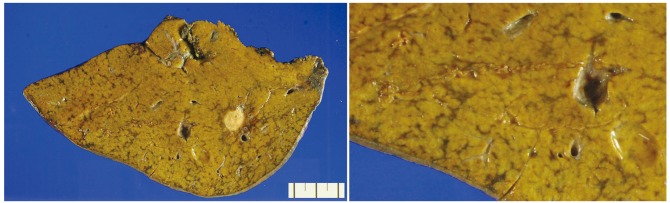
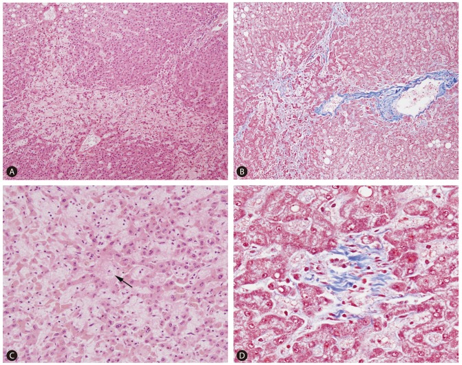
On gross examination, the resected liver showed 2 yellowish-white nodules corresponding to the metastatic lesions, and the background liver showed a distinct mottled appearance, with areas of congestion alternating with relatively normal-appearing hepatic parenchyme (
Fig. 1). Microscopically, the nodules were consistent with metastatic adenocarcinomas from the colorectum. The non-tumorous liver parenchyme showed diffuse sinusoidal dilatation and congestion with extravasation of red blood cells, which predominantly affected the centrilobular zones. Hepatocytic plate disruption was seen in areas of severe sinusoidal congestion, and parenchymal extinction lesions (PELs) - defined as the approximation of hepatic vein remnants and portal tracts with loss of intervening hepatocytes - were noted. Masson's trichrome stain high-lighted centrilobular venular fibrosis and perisinusoidal fibrosis, and fibrous occlusion of small terminal hepatic venules was seen (
Fig. 2). Macrovesicular and microvesicular steatosis was seen in less than 33% of hepatocytes. Hepatocellular ballooning was not prominent, and there was no significant lobular or portal inflammation. There was no significant cholestasis or ductular reaction.
DISCUSSION
Oxaliplatin-based chemotherapeutic regimens such as FOLFOX (5-fluorouracil (5-FU), leucovorin and oxaliplatin) and XELOX (capecitabine and oxaliplatin) are commonly used for patients with colorectal cancer liver metastases - preoperative chemotherapy has been shown to reduce the size of the hepatic metastases, subsequently rendering the tumor more amenable to curative resection.
3,
4 However, while these drugs have demonstrated impressive results in the treatment of colorectal cancers, they have also been associated with various degrees of hepatotoxicity. 5-FU has been associated with hepatic steatosis, possibly mediated by excessive production of reactive oxygen species resulting in the accumulation of lipid vesicles in hepatocytes,
3,
7,
8 and an association between irinotecan and steatohepatitis has been reported.
8,
9 Oxaliplatin has been frequently associated with SOS. SOS has been described in association with drugs including oxaliplatin, azathioprine, cysteamine, dacarbazine, dactinomycin, carmustine (BCNU), 6-mercaptopurine, 6-thioguanine, busulfan, dimethyl busulfan, cytosine arabinoside, cyclophosphamide, indicine-N-oxide, mustine-HCl, doxorubicin, urethane, vincristine, mitomycin-C, etoposide, arsenic, thorium dioxide (Thorotrast), and intra-arterial flurodeoxyuridine.
10 SOS related to oxaliplatin administration was first described by Rubbia-Brandt et al,
5 and is characterized by the following histologic findings: sinusoidal dilatation and congestion, centrilobular vein fibrosis and obstruction, perisinusoidal fibrosis, necrosis of pericentral hepatocytes, PELs, hepatocyte plate disruption, and nodular regenerative hyperplasia.
2,
3,
4,
6,
10,
11 These changes are generally irregularly distributed within the hepatic parenchyme; however, if the changes are more extensive it is possible to recognize SOS on gross examination, where the liver appears diffusely "nodular" due to alternating areas of congestion/hemorrhage with relatively normal-looking areas.
Although the pathogenesis of chemotherapy-induced SOS is still unclear, it has been suggested that initial toxic damage to sinusoidal endothelial cells results in the disruption of the sinusoidal wall, resulting in marked sinusoidal dilatation and extravasation of erythrocytes into the Disse's spaces through the discontinuities in the endothelial lining. In addition, activation of hepatic stellate cells results in the deposition of collagen matrix in the perisinusoidal spaces and centrilobular vein.
3,
5 The degree of SOS has been suggested to be time-and dose-dependent- the extent of sinusoidal dilatation has been shown to be significantly higher in livers exposed to higher numbers of chemotherapy cycles.
3,
11 Interestingly, Overman et al.
12 demonstrated that oxaliplatin-increased SOS was associated with portal hypertension, splenomegaly and subsequent thrombocytopenia, and suggested that increased spleen size could correlate with increasing grade of hepatic sinusoidal injury.
12
The relationship between preoperative chemotherapy-related liver pathology and postoperative outcome is still controversial. Some studies have demonstrated a lack of significant association between chemotherapy-associated hepatopathy and postoperative outcome.
6,
9,
13 On the other hand, preoperative chemotherapy was significantly correlated with increased postoperative morbidity and abnormal liver function tests such as AST, ALT, and total bilirubin in another recent study, suggesting that preoperative chemotherapy may lead to some derangement of liver function.
3 There is limited data regarding the postoperative outcome in relation to SOS following oxaliplatin-based treatment, but an association between the presence of SOS and increased postoperative morbidity has been suggested: sinusoidal injury has been associated with a significantly higher complication rate and poorer liver functional reserve among patients undergoing a major hepatectomy, suggesting the importance of careful surgical candidate selection in patients previously exposed to oxaliplatin-based treatment.
11
FOOTNOTES
-
The authors have no conflicts to disclose.
Abbreviations
Parenchymal extinction lesion
sinusoidal obstruction syndrome
REFERENCES
- 1. DeLeve LD, Shulman HM, McDonald GB. Toxic injury to hepatic sinusoids: sinusoidal obstruction syndrome (veno-occlusive disease). Semin Liver Dis 2002;22:27-42. 11928077.
- 2. Kandutsch S, Klinger M, Hacker S, Wrba F, Gruenberger B, Gruenberger T. Patterns of hepatotoxicity after chemotherapy for colorectal cancer liver metastases. Eur J Surg Oncol 2008;34:1231-1236. 18272318.
- 3. Nam SJ, Cho JY, Lee HS, Choe G, Jang JJ, Yoon YS, et al. Chemotherapy-associated hepatopathy in korean colorectal cancer liver metastasis patients: oxaliplatin-based chemotherapy and sinusoidal injury. Korean J Pathol 2012;46:22-29. 23109974.
- 4. Robinson SM, Wilson CH, Burt AD, Manas DM, White SA. Chemotherapy-associated liver injury in patients with colorectal liver metastases: a systematic review and meta-analysis. Ann Surg Oncol 2012;19:4287-4299. 22766981.
- 5. Rubbia-Brandt L, Audard V, Sartoretti P, Roth AD, Brezault C, Le Charpentier M, et al. Severe hepatic sinusoidal obstruction associated with oxaliplatin-based chemotherapy in patients with metastatic colorectal cancer. Ann Oncol 2004;15:460-466. 14998849.
- 6. Ryan P, Nanji S, Pollett A, Moore M, Moulton CA, Gallinger S, et al. Chemotherapy-induced liver injury in metastatic colorectal cancer: semiquantitative histologic analysis of 334 resected liver specimens shows that vascular injury but not steatohepatitis is associated with preoperative chemotherapy. Am J Surg Pathol 2010;34:784-791. 20421779.
- 7. Khan AZ, Morris-Stiff G, Makuuchi M. Patterns of chemotherapy-induced hepatic injury and their implications for patients undergoing liver resection for colorectal liver metastases. J Hepatobiliary Pancreat Surg 2009;16:137-144. 19093069.
- 8. Morris-Stiff G, Tan YM, Vauthey JN. Hepatic complications following preoperative chemotherapy with oxaliplatin or irinotecan for hepatic colorectal metastases. Eur J Surg Oncol 2008;34:609-614. 17764887.
- 9. Vauthey JN, Pawlik TM, Ribero D, Wu TT, Zorzi D, Hoff PM, et al. Chemotherapy regimen predicts steatohepatitis and an increase in 90-day mortality after surgery for hepatic colorectal metastases. J Clin Oncol 2006;24:2065-2072. 16648507.
- 10. Wanless IR. Vascular disorders of the liver. In: Saxena R, ed. Practical hepatic pathology - a diagnostic approach. Philadelphia: Saunders; 2011. p. 443-454.
- 11. Nakano H, Oussoultzoglou E, Rosso E, Casnedi S, Chenard-Neu MP, Dufour P, et al. Sinusoidal injury increases morbidity after major hepatectomy in patients with colorectal liver metastases receiving preoperative chemotherapy. Ann Surg 2008;247:118-124. 18156931.
- 12. Overman MJ, Maru DM, Charnsangavej C, Loyer EM, Wang H, Pathak P, et al. Oxaliplatin-mediated increase in spleen size as a biomarker for the development of hepatic sinusoidal injury. J Clin Oncol 2010;28:2549-2555. 20406923.
- 13. Nordlinger B, Sorbye H, Glimelius B, Poston GJ, Schlag PM, Rougier P, et al. Perioperative chemotherapy with FOLFOX4 and surgery versus surgery alone for resectable liver metastases from colorectal cancer (EORTC Intergroup trial 40983): a randomised controlled trial. Lancet 2008;371:1007-1016. 18358928.
Figure 1Macroscopic finding of sinusoidal obstruction syndrome. The non-neoplastic hepatic parenchyme is diffusely mottled, with geographic areas of congestion alternating with relatively normal-appearing liver.
Figure 2Representative microscopic findings of sinusoidal obstruction syndrome (A-D). Patchy perivenular sinusoidal dilatation and congestion with hepatocyte plate disruption (A ×100 magnification). Approximation of portal structures and hepatic vein with loss of intervening hepatocytes, and perivenular and perisinusoidal fibrosis (B ×100 magnification, Masson's trichrome stain). Sinusoidal dilatation and congestion with atrophy and disruption of hepatocyte plates (C ×200 magnification). An obliterated terminal hepatic venule is seen in the center (arrow). Masson's trichrome stain demonstrating fibrous obliteration of a terminal hepatic venule (D ×400 magnification).
Citations
Citations to this article as recorded by

- Imaging of hepato-pancreato-biliary emergencies in patients with cancer
Ankush Jajodia, Philippe Soyer, Maxime Barat, Michael N. Patlas
Diagnostic and Interventional Imaging.2024; 105(2): 47. CrossRef - Blue Liver
Kiyoshi Takemoto, Saiho Ko, Kazuaki Atagi
The American Journal of the Medical Sciences.2024; 368(4): e43. CrossRef - Pancreaticoduodenectomy with combined hepatic artery and portal vein resection after laparoscopic division of pancreaticosplenic ligament due to FOLFIRINOX-induced hepatic toxicity related secondary hypersplenism
Ji Su Kim, Jeong Youp Park, Munseok Choi, Dong Jin Joo, Ho Kyoung Hwang, Woo Jung Lee, Chang Moo Kang
Annals of Hepato-Biliary-Pancreatic Surgery.2021; 25(2): 307. CrossRef - Implications of Breast Cancer Chemotherapy-Induced Inflammation on the Gut, Liver, and Central Nervous System
Taurean Brown, DeLawrence Sykes, Antiño R. Allen
Biomedicines.2021; 9(2): 189. CrossRef - Association of albumin–bilirubin score in patients with colorectal cancer receiving later-line chemotherapy with regorafenib
Daichi Watanabe, Hironori Fujii, Yunami Yamada, Nobuhisa Matsuhashi, Akitaka Makiyama, Hirotoshi Iihara, Takao Takahashi, Shigeru Kiyama, Ryo Kobayashi, Kazuhiro Yoshida, Akio Suzuki
International Journal of Clinical Oncology.2021; 26(7): 1257. CrossRef - Hepatic sinusoidal obstruction syndrome/veno-occlusive disease after hematopoietic cell transplantation: historical and current considerations in Korea
Jae-Ho Yoon, Chul Won Choi, Jong-Ho Won
The Korean Journal of Internal Medicine.2021; 36(6): 1261. CrossRef - Imaging of Colorectal Liver Metastases: New Developments and Pending Issues
Matteo Renzulli, Alfredo Clemente, Anna Maria Ierardi, Irene Pettinari, Francesco Tovoli, Stefano Brocchi, Giuliano Peta, Salvatore Cappabianca, Gianpaolo Carrafiello, Rita Golfieri
Cancers.2020; 12(1): 151. CrossRef - Peripheral hepatic sinusoidal obstruction syndrome due to oxaliplatin‐based chemotherapy
Shinichiro Kobayashi, Sadayuki Okudaira, Kazuma Kobayashi, Kengo Kanetaka, Susumu Eguchi
Clinical Case Reports.2019; 7(2): 394. CrossRef - The Protective Effect of Naringenin on Oxaliplatin-Induced Genotoxicity in Mice
Majid A. Ganaie, Basit L. Jan, Tajdar H. Khan, Khalid M. Alharthy, Ishfaq A. Sheikh
Chemical and Pharmaceutical Bulletin.2019; 67(5): 433. CrossRef - Vitamin E protects human lymphocytes from genotoxicity induced by oxaliplatin
Mohammad A.Y. Alqudah, Fahmi Y. Al-Ashwal, Karem H. Alzoubi, Mohammad Alkhatatbeh, Omar Khabour
Drug and Chemical Toxicology.2018; 41(3): 281. CrossRef - Imaging features of hepatic arterial and venous flow abnormalities
Abigail Mills, Vincent Mellnick
Clinical Liver Disease.2018; 11(1): 27. CrossRef - Liquiritigenin and liquiritin alleviated MCT-induced HSOS by activating Nrf2 antioxidative defense system
Zhenlin Huang, Yuchen Sheng, Minwei Chen, Zhanxia Hao, Feifei Hu, Lili Ji
Toxicology and Applied Pharmacology.2018; 355: 18. CrossRef - Commitment of Scaffold Proteins in the Onco-Biology of Human Colorectal Cancer and Liver Metastases after Oxaliplatin-Based Chemotherapy
Deborah Rotoli, Manuel Morales, Julio Ávila, María Maeso, María García, Ali Mobasheri, Pablo Martín-Vasallo
International Journal of Molecular Sciences.2017; 18(4): 891. CrossRef - Oral siRNA Delivery to Treat Colorectal Liver Metastases
Sung Hun Kang, Vishnu Revuri, Sang-Joon Lee, Sungpil Cho, In-Kyu Park, Kwang Jae Cho, Woo Kyun Bae, Yong-kyu Lee
ACS Nano.2017; 11(10): 10417. CrossRef - Target hepatic artery regional chemotherapy and bevacizumab perfusion in liver metastatic colorectal cancer after failure of first-line or second-line systemic chemotherapy
Hui Chen, Ji Zhang, Guang Cao, Peng Liu, Haifeng Xu, Xiaodong Wang, Xu Zhu, Song Gao, Jianhai Guo, Linzhong Zhu, Pengjun Zhang
Anti-Cancer Drugs.2016; 27(2): 118. CrossRef - Chlorogenic acid suppresses monocrotaline-induced sinusoidal obstruction syndrome: The potential contribution of NFκB, Egr1, Nrf2, MAPKs and PI3K signals
Zhiyong Zheng, Liang Shi, Yuchen Sheng, Jiaqi Zhang, Bin Lu, Lili Ji
Environmental Toxicology and Pharmacology.2016; 46: 80. CrossRef - Cancer therapy related complications in the liver, pancreas, and biliary system: an imaging perspective
Danny Ngo, Jemianne Bautista Jia, Christopher S. Green, Anjalie T. Gulati, Chandana Lall
Insights into Imaging.2015; 6(6): 665. CrossRef - Management intra- und postoperativer Blutungen in der Leberchirurgie
R. Sucher, D. Seehofer, J. Pratschke
Der Chirurg.2015; 86(2): 114. CrossRef - Imaging of Fluid in Cancer Patients Treated With Systemic Therapy: Chemotherapy, Molecular Targeted Therapy, and Hematopoietic Stem Cell Transplantation
Abhishek R. Keraliya, Michael H. Rosenthal, Katherine M. Krajewski, Jyothi P. Jagannathan, Atul B. Shinagare, Sree Harsha Tirumani, Nikhil H. Ramaiya
American Journal of Roentgenology.2015; 205(4): 709. CrossRef - Capecitabine/oxaliplatin
Reactions Weekly.2015; 1548(1): 61. CrossRef - Chemotherapy induced liver abnormalities: an imaging perspective
Ankush Sharma, Roozbeh Houshyar, Priya Bhosale, Joon-Il Choi, Rajesh Gulati, Chandana Lall
Clinical and Molecular Hepatology.2014; 20(3): 317. CrossRef