Clin Mol Hepatol > Volume 26(2); 2020 > Article
Chen, Wang, and Xiao: Regulated differentiation of stem cells into an artificial 3D liver as a transplantable source

ABSTRACT

End-stage liver disease is one of the leading causes of death around the world. Since insufficient sources of transplantable liver and possible immune rejection severely hinder the wide application of conventional liver transplantation therapy, artificial three-dimensional (3D) liver culture and assembly from stem cells have become a new hope for patients with end-stage liver diseases, such as cirrhosis and liver cancer. However, the induced differentiation of single-layer or 3D-structured hepatocytes from stem cells cannot physiologically support essential liver functions due to the lack of formation of blood vessels, immune regulation, storage of vitamins, and other vital hepatic activities. Thus, there is emerging evidence showing that 3D organogenesis of artificial vascularized liver tissue from combined hepatic cell types derived from differentiated stem cells is practical for the treatment of end-stage liver diseases. The optimization of novel biomaterials, such as decellularized matrices and natural macromolecules, also strongly supports the organogenesis of 3D tissue with the desired complex structure. This review summarizes new research updates on novel differentiation protocols of stem cell-derived major hepatic cell types and the application of new supportive biomaterials. Future biological and clinical challenges of this concept are also discussed.

INTRODUCTION

In the clinical context, chronic liver disease is a process in which the liver undergoes progressive destruction and regeneration, which lead to fibrosis and cirrhosis. It is one of the major causes of mortality worldwide. In 2016, chronic liver disease and cirrhosis caused a total of 40,545 deaths, in which alcohol-related liver disease induced nearly half of the deaths [1]. Since damage in the liver often induces inflammation, oxidative stress, cellular necrosis and apoptosis, which causes tissue scarring and fibrosis if occurring repeatedly, developing therapies that can retard or reverse those pathological processes is quite important [2]. When liver disease progresses to the end stage, it becomes irreversible and liver failure is inevitable [3]. At this point, liver transplantation is the only clinical option. However, the lack of transplantable livers and the possibility of immune rejection after the transplant seriously hinders its wide application [4]. In addition, the maximal cold preservation duration of the human liver organ soaked in University of Wisconsin solution before a clinical transplantation operation is only 6–12 hours [5]. Developing novel supercooling techniques to extend this duration to 24–36 hours will dramatically enlarge the donation territory, as well as reduce logistical pressures and facilitate recipient preparation.
To solve the problems hindering successful clinical liver transplants, stem cell medicine provides a brand new solution, particularly those stem cells with the potential to differentiate into liver cells that can then be transplanted directly. For example, a large number of studies have proved that the transplantation of differentiated hepatocytes from human adipose derived stem cells, human amniotic epithelial stem cells, and inducible pluripotent stem cells (iPSCs) effectively ameliorated the complications induced by acute liver failure, including elevated serum aminotransferases, hepatic inflammation, and cell death [6-9]. Stem cell therapy was also reported to be successful in other kinds of chronic/end-stage liver diseases, including nonalcoholic steatohepatitis [10], Wilson’s disease [11], cirrhosis [12], and hepatocellular carcinoma [13]. However, mechanistic studies found that the life support and hepatic protective effects of exogenous stem cells, including mesenchymal stem cells (MSCs) and iPSCs, were primarily from their paracrine actions instead of from direct hepatic occupation [14]. Only hepatic progenitor cells were able to completely and functionally reconstitute a liver that had suffered otherwise lethal damage [15].
Although strategies with differentiated hepatocytes and naïve stem cells demonstrated massive success in the past decades, single cell type-based injection or “in situ transplantation” cannot meet the majority of clinical requirements, which need physiologically functional hepatic tissue to ameliorate damage and support normal liver functions within a short duration. To solve this problem, scientists began to use a complicated but fascinating method – constructing artificial organs from stem cells in three-dimensional (3D) culture. In the current review, we focus on the research updates on liver organogenesis from stem cells, with emphases on the differentiation protocol, biomaterial support, and self-condensation mechanisms.

3D ORGANOGENESIS FROM STEM CELLS

Over the past decade, significant progress has been made in controlling cellular differentiation in stem cell research. For example, it is now possible to force MSCs and iPSCs to differentiate into a large number of specific somatic cell lineages by mimicking the signals presented during embryogenesis. Very importantly, several studies have demonstrated that stem cells have the ability to self-organize into a functional tissue by scattering various somatic cells throughout the tissue [16-19]. For example, Takebe et al. [17] generated a vascularized and functional human liver from human iPSCs by transplanting liver buds created in vitro. Such liver buds were created from a mixture of human iPSCs, human MSCs, and human umbilical vein endothelial cells (HUVECs). The cells then underwent self-organization into a 3D liver bud by recapitulating the organogenetic interactions between the endothelial and mesenchymal cells [19]. The authors tested the hepatic functions of the liver buds both in vitro and in a murine liver failure model, which exhibited satisfactory outcomes. They then expanded this strategy to even more vascularized artificial organs, including kidney, pancreas, intestines, heart, lungs, and brain. It was found that mesenchyme-driven self-condensation on a soft matrix is crucial for organ bud generation [18]. Although those in vitro-grown complex tissue architectures have limited size and survival duration, the ideas raised by this Japanese group extensively inspired other regenerative medicine researchers and clinicians to consider that a mixture of hepatic cell types that are necessary for organ development and self-condensation (e.g., hepatocytes, stellate cells, sinusoidal endothelial cells, and bile duct epithelial cells), together with guides from undifferentiated stem cells, is a promising strategy to generate a clinically transplantable liver or other organ.

DIFFERENTIATION INTO HEPATIC CELL TYPES

Hepatocytes

Hepatocytes are the major cell type of the liver and account for approximately 80% of the hepatic volume and quantity. They are a highly differentiated parenchymal cell type. There are more than 2.5 billion hepatocytes in an adult human liver, and a hepatic lobule is made up of 5 million hepatocytes. Active hepatocyte metabolism can synthesize human essential blood coagulation factors, aliphatic acid, cholesterol, and phospholipids; hepatocytes are also able to store glycogen, proteins, lipids, and vitamins to fulfill the body’s needs.
In the liver, hepatocytes are the very basic cells that support its essential physiological functions. Over the past decade, several studies have introduced protocols to induce hepatocyte-differentiation in stem cells; most methods use chemical and conditioned medium stimulations or coculture with other cell types to expedite hepatic differentiation from human bone marrow mesenchymal stem cells, human umbilical cord mesenchymal stem cells, and human iPSCs [20-23]. Induced hepatocyte-like cells (i-Hep) are shown to possess most of the normal hepatocyte functions (including the secretion of aminotransferases, albumin, and α-fetoprotein), functional biotransformation systems, and the ability to engraft, integrate, and proliferate in an injured liver. Nie et al. [24] used human iPSC to generate a functional liver organoid, which exhibited stronger hepatic functions than human iPSC-derived hepatocyte-like cells (or i-Hep). They found that the functional liver organoid was more susceptible to hepatitis B virus (HBV) infection and could maintain HBV propagation and produce the infectious virus for a prolonged duration, thus causing the hepatic dysfunction of the liver organoid via the down-regulation of hepatic gene expression, induced release of early acute liver failure markers, and altered hepatic ultrastructure. The liver organoid may provide a promising individualized infection model for the development of individualized treatment for hepatitis [24]. Different stem cell types require different concentrations of inducing reagents, including hepatocyte growth factor (HGF), fibroblast growth factor (FGF), oncostatin M (OSM), epidermal growth factor (EGF), and bone morphogenetic protein [25]. Currently, one can even conveniently purchase a commercial one-step direct hepatocyte induction kit.
Although still in the primary stages, a number of studies have tried to induce and culture differentiated hepatocytes in a 3D manner in vitro. The primary interest in developing 3D-structured hepatocytes is because although the 2D in vitro culture of hepatocytes is currently mature, 2D-cultures show a reduction in major liver functions, such as a decreased secretion of albumin and impaired phase I and II enzymatic detoxification abilities [26]. The application of extracellular matrix (ECM) is a major solution for these problems. Thus far, the most common strategy is the sandwich structure in which hepatocytes are placed between two layers of ECM. This model has been proven to provide better hepatocyte cellular functions than 2D monolayer culture conditions since it promotes a polygonal hepatocyte morphology and extended contact surfaces between the cells and the matrix [27].

Hepatic stellate cells (HSCs)

HSCs are the major nonparenchymal cells of the liver. Under physiological conditions, HSCs are in a quiescent state in which their main function is to store and transport vitamin A [28]. When the liver is damaged, under the actions of inflammatory cytokines and stress factors, HSCs change into an activated state (a myofibroblastic phenotype) characterized by increased proliferation, contractility, and chemotaxis. The activation of HSCs will promote the secretion of the ECM involved in liver injury repair. Thus, HSCs play an important role in the occurrence and development of various liver diseases. Moreover, through their interaction with other liver cell types, HSCs are also involved in liver regeneration and differentiation [29].
Research on HSC formation from stem cells is scarce. The main reason for this is that the embryonic origin of HSCs is yet unresolved, with hypotheses of mesenchymal and endodermal origins [30]. Baba et al. [31] proved that in a murine model, HSCs are from the bone marrow since they administered bone marrow cells from green fluorescent protein (GFP) transgenic mice to age-matched mice, and found GFP-positive HSCs in the recipient livers. However, an analytical study of cell lineage demonstrated that HSCs are derived from the mesoderm during liver development; in particular, they are derived from the mesothelium (consisting of mesothelial cells and submesothelial cells) which migrate inward from the liver surface to form HSCs and perivascular mesenchymal cells [32]. Other studies claimed that HSCs are important for the association with hematopoietic sites in the fetal rat liver. HSCs can fulfill some of the functions of MSCs, such as adipocytes or osteocytes differentiation [33,34]. Thus far, only Coll et al. [35] reported that they could obtain HSC-like cells from newborn male fibroblastsderived human iPSC by mimicking embryonic development; those cells closely resemble primary human HSCs at the transcriptional, cellular, and functional levels and possess a gene expression profile between that of quiescent and activated HSCs.

Liver sinusoidal endothelial cells (LSECs)

LSECs consist of the majority of liver nonparenchymal cells (~70% of the total liver nonparenchymal cells). They are functionally unique because of the high activity of receptor-mediated endocytosis, which enables them to clear colloids and soluble waste macromolecules from circulation [36]. LSECs have long been noted to contribute to liver regeneration after liver injury. That is, there is crosstalk between HSCs and LSECs when the activation of HSCs and the formation of fibrosis occur. Healthy LSECs are able to suppress the activation of HSCs and also inactivate already-activated HSCs [37]. Conversely, in the healthy liver, hepatocytes and HSCs can work together to maintain the homeostasis of LSECs through the release of vascular endothelial growth factor (VEGF) [38]. When the Kupffer cells (KCs)’ phagocytic function is damaged, LSECs will remove materials with diameters greater than 1 μm. Furthermore, LSECs take in viruses and submit antigens to T cells. Additionally, they participate in the local immune regulation in the liver and may also play an important role in immune tolerance [39].
Reports of direct differentiation from stem cells to LSECs are extremely limited. Du et al. [40] demonstrated a novel two-step method for that process, that is, a 7-day first stage from human iPSCs that are generated by reprogramming human foreskin fibroblasts to the mesodermal lineage and a 7-day second stage from mesodermal cells to hepatic endothelial cells. The final LSEC-like cells are positive for expressions of endothelial-specific markers such as CD-31 and VE-cadherin [40]. Vunjak-Novakovic et al. [41] claimed that they established a culture condition to promote the differentiation from multiple iPSC cell lines to endothelial cells that coexpress cell surface markers and functional properties (e.g., endothelial nitric oxide synthase production) with an efficiency of approximately 10–40%. In addition, Narmada et al. [42] declared that they had formed endothelial cells from human iPSC that had been derived from IMR90 fibroblasts and BJ fibroblasts by using a chemically defined hepatocyte growth medium (HGM); those cells expressed PECAM-1 and CDH5, could form spontaneous tube structures that represent mature endothelial markers, possessed endothelial nitric oxide synthase, and were capable of taking up acetylated low-density lipoprotein.

Bile duct epithelial cells (BDECs)

BDECs are a kind of distinctive epithelial cell coating on the liver inner tube surface. They are of great importance in bile duct contraction, bile secretion, and liver water electrolyte transport. In addition, BDECs can also mediate the synthesis and secretion of IgA/IgM, cytokines, chemokines, and adhesion molecules involved in immunity, cell interaction, and signal transduction [43].
A study described a differentiation procedure from embryonic stem cells (ESCs) to BDECs on the basis of induced hepatocytes. The authors coincubated the ESCs with HGF/scatter factor, EGF, and transforming growth factor α (TGF-α) in the presence of a new HGM. In addition to the expected morphology, the differentiated BDECs also expressed cytokeratin 19, acidic FGF, and TGF-α, but did not express the hepatocyte markers including albumin and cytochrome P450 IIB1 [44]. Another study confirmed that dexamethasone and transforming growth factor β (TGF-β) contribute to differentiation into BDECs; these compounds are closely connected with the Wnt/β-catenin associated protein, and the Notch and TGF-β signaling transduction pathways [45]
Sampaziotis et al. [46,47] from the University of Cambridge reported a novel and serum-free method for the direct differentiation of cholangiocytes from human skin fibroblasts and peripheral blood derived iPSCs. By using several combinations of recombinant growth factors and inhibitors at different stages, they formed definitive endoderms, foregut progenitors, hepatoblasts, and cholangiocyte progenitors from iPSCs in sequence. After a 26-day differentiation process, the final cholangiocyte-like cells were capable of showing the functional characteristics of normal cholangiocytes, including alkaline phosphatase/γ-glutamy-ltranspeptidase activity, transfer of bile acids, and responses to secretin, somatostatin, and VEGF. The derived cells were also useful in establishing some common biliary disease models (e.g., Alagille syndrome, polycystic liver disease, and cystic fibrosis-associated cholangiopathy) [46,47].

Kupffer cells (KCs)

KCs are hepatic local macrophages which are located on the inner surface of the sinus hepaticus. They are a critical component of the mononuclear phagocytic system, which takes part in the response to pathogens. KCs can remove foreign antigens, antigen-antibody complexes, cell debris, and other substances in the liver and circulation partly through the activation of nuclear factor kappaB and the secretion of a variety of cytokines [48]. The abnormal activation and regulation of KCs contribute to nearly all types of liver diseases, such as acute liver failure, nonalcoholic fatty liver disease, alcohol-related liver disease, genetic liver diseases, cirrhosis, and liver cancer [49].
Although a number of studies have successfully established methods to generate macrophage or dendritic cell from human PSCs [50,51], no report described the differentiation protocol from stem cells to KCs until 2018. Part of the reason is that the derived macrophage is able to migrate into the liver to form KCs and pit cells (intrahepatic lymphocytes). In addition, in the process of establishing an artificial 3D liver, KCs and pit cells are not necessary because 1) they are expected to migrate into the engineered liver grafts from the bone marrow and the circulatory system of the recipients upon transplantation [52], 2) the functions of the assembled LSECs are similar to those of KCs [53], and 3) KCs may secrete nitric oxide to increase their own apoptosis, leading to acute rejection after liver transplantation. Additionally, KCs will induce apoptosis and differentiation of T-cells through the Fas-FasL apoptosis pathway [54]. Thus far, only Tasnim et al. [55] demonstrated that they could obtain mature KCs from human IMR90 fibroblasts derived iPSCs (iKCs). By using a different culture medium and several combinations of recombinant growth factors and inhibitors at different stages, they generated KCs that expressed the macrophage markers CD11, CD14, CD68, CD163, CD32 at 0.3–5 folds of those from primary adult human KCs (pKCs) and KC-specific CLEC-4F, ID1, and ID3. Moreover, iKCs phagocytosed and secreted IL-6 and TNF-α upon stimulation at levels similar to pKCs but different from nonliver macrophages [55]. Although not necessary for 3D liver assembly, KCs should be included in the 3D liver for disease modeling and drug discovery in vitro. The reported differentiation protocols of the above-mentioned hepatic cell types are listed in Table 1. Key markers for differentiated liver cell types were summarized in Figure 1.

ASSEMBLY OF TRANSPLANTABLE 3D LIVER TISSUE

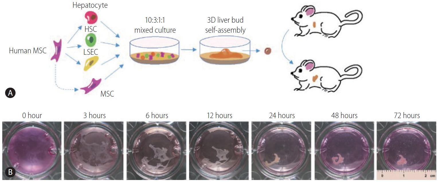
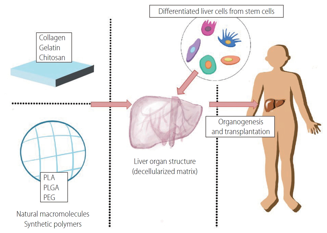
In previous decades, numerous studies have focused on the construction of liver tissue in a 2D planar environment. However, such a strategy is challenging because the actual liver tissue is 3D. Therefore, to address the limitations of 2D cell cultures and achieve the functional recovery of the liver, a 3D microenvironment which aims to mimic the native environment must be fabricated [56]. Recently, scientists focused on exploring artificial 3D ECM by fabricating a scaffold using particular materials or a matrix, including a decellularized matrix, natural macromolecules, and synthetic biodegradable polymers (listed in Table 2) [57]. The engineered scaffolds can provide a 3D environment for cell attachment and proliferation. Moreover, the scaffolds play a role in regulating cell maturation and function by modification with specific physical and biochemical properties [58,59]. Recently, by transdifferentiating hepatocytes, HSCs, and LSECs from human umbilical cord blood stem cells simultaneously, we obtained a self-assembled functional 3D human liver bud in vitro and transplanted it into a murine model of acute liver failure. The liver bud underwent maturation during injury alleviation, after which the cells exhibited a gene expression profile signature similar to that of adult human livers (Fig. 2) [60].

Decellularized matrix

A decellularized matrix scaffold, which is procured by a decellularization technique that removes parenchymal cells from tissues or organs, can potentially retain the skeletal architecture and functional characteristics of the original tissue [61,62]. Decellularized scaffolds derived from animal organs are now being explored as a promising resource for generating transplantable and functional organs. Lin et al. [63] documented that a rat primary hepatocyte culture in a porcine liver-derived biomatrix for up to 45 days showed better liver-specific functions, such as albumin synthesis, urea production, and P450 1A1 activity, than those on adsorbed collagen cultures. Uygun et al. [64] seeded hepatocytes in a decellularized whole liver tissue. In vitro, the recellularized graft supported liver-specific functions including albumin secretion, urea synthesis and cytochrome P450 expression at levels comparable to a normal liver. When transplanted into rats, the recellularized liver grafts could support hepatocyte survival and function with minimal ischemic damage [64]. Park et al. [65] reported that they reseeded hepatocytes derived from iPSCs that had been generated by porcine ear fibroblasts into decellularized liver scaffolds, using a continuous perfusion system to culture the recellularized liver. When the grafts were transplanted into rats for the short-term, they found that the grafts expressed hepatocyte markers and did not rupture [65]. Furthermore, researchers have found that hepatocyte-like cells derived from human amniotic epithelial, bone marrow, and adipose stromal cells display enhanced functionality when cultured on a decellularized liver substrate [66]. To overcome the limitations due to the shortage of donor organs, Xiang et al. [67] utilized the spleen as the source of a decellularized scaffold for liver regeneration. The decellularized spleen scaffold could support the location and survival of BMSCs within the 3D matrix. Moreover, the subcutaneous implantation of the decellularized scaffold presented good histocompatibility [67]. Another study also proved that a bioartificial liver constructed by a heparin-coated decellularized spleen matrix can exert satisfactory short-term anticoagulant effects and can compensate for a certain degree of liver function [68].
However, the strategy based on a decellularized matrix suffers from limitations including a severe shortage of donor organs, high cost of treatment, and lifetime immune suppression [69].

Natural macromolecules

Natural macromolecules that are derived from ECM have been widely investigated as tissue engineering scaffolds for the culture of liver cells, including proteins such as collagen and gelatin as well as polysaccharides such as chitosan and alginate. These macromolecules have shown good biocompatibility both in vitro and in vivo.
Collagen, especially type I collagen, is the main structural protein in the extracellular space in various connective tissues in animal bodies. The collagen matrix serves to mediate cell growth, differentiation, and survival, and tissue organization. A collagen gel could provide a 3D microenvironment for hepatocytes to sustain high liver-specific functions [70]. Khodabandeh et al. [71] cultured human Wharton’s jelly MSCs in 2D collagen films and 3D collagen scaffolds for 21 days. The cells cultured in the 3D collagen scaffolds showed a significant increase in claudin expression compared to a conventional monolayer culture and the 2D collagen scaffold. Their subsequent study showed that the expression of the hepatocyte markers were increased significantly when administering the liver extract from a 3D matrigel/collagen culture of MSCs [72]. Nakai et al. [73] tested the effect of culturing human iPSCs-derived endoderm cells on a collagen vitrigel membrane; they found collagen vitrigel had promoted the differentiation of cells into functional in vitro cells, as shown by the decrease of immature marker alpha-fetoprotein (AFP) levels and the concomitant increase in expression of mature hepatocyte transcription factors (such as ALB and ASGR1 ) and markers involved in liver functions (such as transporters, cytochrome P450 enzymes, and phase II metabolic enzymes). Moreover, collagen gel could also be used in a hepatocyte entrapment culture as an effective model to interpret drug hepatotoxicity and drug metabolism in vivo [74].
Gelatin is the partially hydrolyzed form of collagen with a chemical composition that is, in many respects, very similar to that of its parent collagen. Recently, gelatin-based scaffolds have been documented to increase cell attachment and differentiation. Vasanthan et al. [75] developed galactose-containing physical cross-linked polyvinyl alcohol/gelatin hydrogel scaffolds via the freeze/thaw technique. Such galactose-containing hydrogels promoted cell-cell and cell-hydrogel interactions, aiding cellular aggregation and leading to the formation of spheroids [75]. Another research found that a gelatin cryogel scaffold successfully promoted the differentiation of human adipose-derived MSCs into hepatocyte-like cells with a higher expression of hepatocyte-specific markers and appropriate functional characteristics, such as increased levels of urea biosynthesis and glycogen storage [76]. On this basis, the researchers optimized the aforementioned scaffold and improved it through laminin incorporation; they found that the cells generated on this scaffold were strikingly similar to a standard hepatocyte cell line in terms of the expression of hepatocyte markers [77]. Moreover, gelatin was also employed to fabricate microcarriers with chitosan for hepatocyte culture [78], and improved the performance of hepatocytes for as long as 2 months [79].
Chitosan is a linear polysaccharide which has good biocompatibility and wide applications in biomedical fields. In the study of Verma et al. [80], HepG2 cells cultured on chitosan cast films formed 3D spheroids and exhibited higher amounts of album and urea synthesis compared to a monolayer culture. Rat hepatocytes seeded on an electrospun scaffold of galactosylated chitosan formed immobile, 3D, flat aggregates, and exhibited superior cell bioactivity with high levels of liver-specific albumin secretion, urea synthesis, and cytochrome P450 enzyme expression [81]. Rajendran et al. [82] established a chitosan nanofiber scaffold via an electrospinning technique combined with a coculture system that consisted of hepatocytes and fibroblasts, and thus developed a new 3D liver model for the maintenance of long-term liver functions. To obtain better biocompatibility, chitosan can be further modified with other macromolecules, such as collagen [83], gelatin [78], alginate [84], and heparin [85]. For example, Fan et al. [86] proved that chitosan/galactosylated hyaluronic acid scaffolds were suitable for improving hepatocytes adhesion and maintaining liver function in a previous work. To adjust the interaction of hepatocytes with the scaffold, they further modified the materials by the incorporation of heparin, which induced the formation of cellular aggregates with enhanced liver-specific metabolic activities and increased cell density [85].
Alginate is a widely employed biomaterial in different areas of drug delivery and tissue engineering because of its good biocompatibility and optimal chemical properties. Its gelation with calcium ions provides calcium-alginate scaffolds with mechanical stability and relative permeability. Hepatocytes could organize in aggregates within calcium-alginate scaffolds, which established real 3D hepatocyte architecture with cell polarity, cell junctions, and microvillus-lined channels with abundant bile canaliculi [87]. Lin et al. [88] demonstrated that bone marrow-derived MSCs cultured in alginate scaffolds for several days displayed several liver-specific markers, such as the expression of genes encoding alpha-fetoprotein, ALB, connexin 32, CYP7A1, and were capable of glycogen storage. To fabricate implantable constructs for liver regeneration, Lau et al [89]. encapsulated murine embryoid bodies (EBs) into a 3D alginate microcavitary hydrogel system for both EB formation and subsequent hepatic lineage differentiation. Urea and albumin production were found to be significantly higher compared to a monolayer culture, demonstrating the beneficial effects of the 3D engineered environment [89]. Shteyer et al. [90] reported alginate scaffolds significantly increased animal survival, prolonged alanine aminotransferase and aspartate transaminase serum levels, and were accompanied by improved liver histology after an extended partial hepatectomy. Another study on acute liver failure in rats showed that umbilical cord stem cell-derived in vitro cells encapsulated in alginate scaffolds effectively attenuated biochemical tests, improved liver cytoarchitecture, increased the expression of albumin, and reduced the level of AFP [91]. Additionally, a new study from Shao et al. [92] constructed carbon nanotube nanocomposites via the layer-by-layer assembly of negatively charged multiwalled carbon nanotubes and positively charged poly (dimethydiallylammonium chloride), and proved that they could provide a potent regulatory signal over neural stem cells.
However, natural macromolecules suffer from a limited range of properties due to difficult processing of bulk material and the possibility of inducing potentially dangerous immune responses when used as xenografts or allografts [93]. Moreover, the extraction and purification of natural macromolecules also leads to significant batch-to-batch variations [94], which can lead to unpredictable cell behavior and consequently impact the performance of cell-based tissue-engineered devices.

SYNTHETIC POLYMERS

Biodegradable synthetic polymers have been attractive candidates for hepatocyte culture in vitro and for making implantable scaffolds in vivo [95]. A major advantage of synthetic polymers over natural macromolecules is the easy control of their physiochemical and biological modifications. Some of the widely used synthetic polymers are polylactic acid, poly(lactide-co-glycolide) (PLGA), and poly(ethyleneglycol) (PEG).
The study of Hanada et al. [96] indicated that a 3D culture using poly-L-lactic acid (PLLA) scaffolds with OSM remarkably enhanced the albumin production and cytochrome P450 1A1/2 capacity during the culture time. This observation was in agreement with those of Jiang et al. [97] and Wang et al. [98], where fetal mouse liver cells were cultured on 3D PLLA scaffolds in the presence of nicotinamide, dimethyl sulfoxide, and OSM. In addition, the in vivo implantation of such engineered liver tissue showed a remarkably higher presence of albumin-positive engrafted cells 15 days after the operation when compared to freshly isolated and cultured cells for 1 day [96]. However, researchers are less likely to use pure PLLA because of poor biocompatibility, and because scaffolds fabricated from single polymers display poor mechanical properties and are not easy to handle. To overcome these limitations, many reports have indicated the possible application of PLLA/poly(£-caprolactone) blends [99,100].
Ranucci and Moghe [101] cultured hepatocytes on porous foams of amorphous PLGA with a wide range of controlled pore-size distributions (approximately 1 to 100 microns) and found that foams with supercellular size voids (~67 microns) promoted the kinetics of 3D aggregation with the most rapid and sustained albumin secretory kinetics. Kasuya et al. [102] developed a 3D stacked culture method using biodegradable PLGA membranes. The hepatocyte layers on the PLGA membranes would reorganize into a 3D stacked structure after PLGA degradation. In the study by Liu et al. [103], ESCs were mixed with Matrigel and immediately seeded in a PLGA scaffold with the appropriate inducing factors. The system enabled the spontaneous formation of spheroids, which differentiated into in vitro cells as evidenced by the expression of liver-specific markers and proteins [103].
However, synthetic biodegradable polymers have poor characteristics for cell culture due to their hydrophobic characteristics and lack of bioactive cues. Therefore, their bioactivities should be improved by chemically grafting bioactive cues [104] and/or modifying the surface with natural macromolecules [105]. Kojima et al. [106] developed avidin-biotin binding systems (ABBS) for the initial attachment of biotinylated HepG2 cells to avidin-adsorbed PLLA surfaces. ABBS assisted HepG2 cells to adhere to a PLLA surface, and the proliferation of these attached cells was comparable with those cultured on collagen. Moreover, the hepatic functions of the attached cells were not significantly changed [106]. Brown et al. [107] modified a nanofibrous PLGA electrospun scaffold with a type I collagen coating. The modified scaffold led to 10-fold greater albumin secretion, 4-fold higher urea synthesis, and elevated transcription of hepatocyte-specific CYP450 genes in primary human hepatocytes compared to the unmodified PLGA scaffolds [107]. Similarly, Bierwolf et al. [108] developed a collagen-coated PLLA electrospun nanofibrous scaffold which provide a good in vitro microenvironment for new tissue regeneration of primary rat hepatocytes. Application of novel materials for liver matrix scaffold construction and organogenesis were illustrated in Figure 3.

CONCLUDING REMARKS

Establishing a transplantable artificial 3D liver is complicated. Numerous studies have proved that a single hepatocyte component may not be able to fulfill the physiologically required liver functions. Thus, a combination of hepatocytes, HSCs, BDECs, and LSECs is proposed to form vascularized liver tissues providing blood circulation and nutrient supply. Existing researches have shown several ways to force stem cells to differentiate into those kinds of cells, with the proper physical supports from novel biomaterials. Indeed, there are a number of problems that need to be overcome, such as 1) ensuring that the differentiation process occurs properly and completely, 2) constructing 3D biomaterials with proper hepatic structures and with controllable release of growth factors for the self-formation of the liver tissue, and 3) avoiding possible tumorigenicity. Addressing these concerns will definitely promote the basic and clinical development of novel strategies for treating various liver diseases.

ACKNOWLEDGMENTS

This work was supported by the Major Science and Technology Projects of Guangdong Province (No. 2015B020225005) and the National Natural Science Foundation of China (No. 81970515, 81873573, 81800525).

FOOTNOTES

Authors’ contribution
All of the authors were responsible for the interpretation of data and drafting and critically revising the manuscript for important intellectual content.
Conflicts of Interest: The authors have no conflicts to disclose.

Figure 1.
Key functional markers of liver cell types differentiated from stem cells (e.g., embryonic stem cells, mesenchymal stem cells, and inducible pluripotent stem cells). Major liver cell types, including hepatocytes, Kupffer cells, hepatic stellate cells, liver sinusoidal endothelial cells, and cholangiocytes can be reprogrammed from various stem cells using defined medium and chemical compounds during in vitro culture. Key functional markers can be used to validate the successful reprogramming of each cell type. ALB, albumin; HNF4α, hepatocyte nuclear factor 4 α; CK, cytokeratin; AFP, α-fetoprotein; AAT, α-1 antitrypsin; CYP, cytochrome P family enzymes; PECAM, platelet endothelial cell adhesion molecule-1; eNOS, endothelial nitric oxide synthase; ESCs, embryonic stem cells; MSCs, mesenchymal stem cells; iPSCs, inducible pluripotent stem cells; ALCAM, activated leukocyte cell adhesion molecule; CRBP, cellular retinol-binding protein; COL1α1, collagen type 1 α 1; GFAP, glial fibrillary acidic protein; RELN, reelin; PCDH7, protocadherin-7; aFGF, acidic fibroblast growth factor.

cmh-2019-0022nf1.jpg
Figure 2.
Human 3D liver bud assembly from human umbilical cord blood stem cells (hUCBSCs). (A) Schematic representation of our strategy to assemble human 3D liver buds from naive MSC and MSC-derived hepatocytes, HSC-like cells, and LSEC-like cells. (B) The time-lapse representative images of the self-assembly process. This data and description have been published by Li et al. [60] MSC, mesenchymal stem cell; HSC, hepatic stellate cell; LSEC, liver sinusoidal endothelial cell.

cmh-2019-0022nf2.jpg
Figure 3.
Assembly of transplantable 3D liver tissue. Natural macromolecules and synthetic polymers can be used to construct a decellularized matrix scaffold. Differentiated liver cells (hepatocytes, Kupffer cells, hepatic stellate cells, liver sinusoidal endothelial cells, and cholangiocytes) are then mixed and transplanted into the matrix scaffold to allow organogenesis for possible transplantation. PLA, polylactic acid; PLGA, poly(lactide-co-glycolide); PEG, poly(ethyleneglycol).

cmh-2019-0022nf3.jpg
Table 1.
Differentiation-induction methods of major liver cell types from stem cells
Stem cell type Species Brief protocol Markers Reference
Hepatocytes
ESCs Human 1st stage: RPMI+B27 with activin and NaB for 5 days; 2nd stage: Knockout DMEM + 20% SR with DMSO for 7 days; 3rd stage: CL15 with HGF and oncostatin M for 7 days Expression of HNF4α, ALB, FoxA2, TTR, A1AT, and AFP [109]
MSCs Human 1st stage: DMEM/F-12 with HGF, EGF, ITS and dexamethasone for 2 weeks; 2nd stage: DMEM/F-12 with HGF, EGF, ITS, dexamethasone and oncostatin M for 2 weeks Expression of CK-18, ALB and AFP [6]
ESCs and iPSCs Human 1st stage: RPMI+B27 with activin A for 5 days; 2nd stage: RPMI+B27 with BMP-4 and FGF2 for 5 days (20% CO2); 3rd stage: RPMI+B27 with BMP-4 and FGF2 for 5 days; 4th stage: RPMI+B27 with HGF; 5th stage: HCM with oncostatin M Secretion of albumin, expression of FOXA2, HNF4α and AFP [110]
ESCs and iPSCs Human 1st stage: RPMI+B27 with CHIR99021 for 1 day and removal of CHIR for another day; 2nd stage: KOSR with DMSO for 5 days; 3rd stage: L15 with Dihexa and dexamethasone for 10 days Expression of albumin, HNF4α, AAT and CYP3A4 [111]
ADSCs Human 1st stage: 10% FBS RPMI-1640 with ATRA for 1day; 2nd stage: serum-free RPMI-1640 with IDE1, CHIR99021, LY294002 for 1 day; 3rd stage: serum-free RPMI-1640 with IDE1, LY294002, LDN-193189, FGF4 for 2 days; 4th stage: serum-free RPMI-1640 with IDE1, LY294002, FGF4 for 1 day; 5th stage: Williams’ E with HGF, FGF4, oncostatin M, dexamethasone, ITS for 5 days Expression of HNF4α, AAT, ALB, TDO2, AFP, CYP3A4, TTR, CK-18. [9]
With capacity for glycogen storage.
PDLSCs Human 1st stage: 2% FBS Knockout DMEM with HGF for 5 days; 2nd stage: Knockout DMEM with oncostatin M, dexamethasone, ITS for 16 days Expression of HNF4α, CK-18, ALB, TAT, TDO, AAT and presence of glycogen storage [20]
ESCs and iPSCs Human 1st stage: mTeSRTM medium with DMSO for 1 day; 2nd stage: RPMI 1640 + B27 with CHIR99021 for 1 day; 3rd stage: RPMI 1640 + B27 alone for 1 day; 4th stage: advance F12 with A83-01, sodium butyrate, DMSO for 5 days; 5th stage: advance F12 with FH1, FPH1, A83-01, dexamethasone, hydrocortisone for 5 days Expression of ALB, AAT, TTR, APOA2, HNF4α, and CYP450 enzymes. Secretion of albumin and capacity for glycogen storage. [22]
iPSCs Porcine 1st stage: RPMI with activin A, Wnt 3a for 1 day; 2nd stage: SFD with activin A, bFGF for 5 days; 3rd stage: SFD with bFGF, BMP4, EGF, HGF for 3 days; 4th stage: SFD with y-secretase inhibitor-X, HGF, OSM, DMSO for 3 days; 5th stage: SFD with HGF, OSM, dexamethasone for 6 days Expression of ALB, HNF1α, CK-18, TFR, CK-8, AFP. Expression of metabolizing phase I phase II enzymes, and phase III transporters. [25]
Hepatic stellate cells
iPSCs Human 1st stage: DMEM with Wnt3a and activin-A for 6 days; 2nd stage: DMEM with bFGF and BMP4 for 4 days; 3rd stage: DMEM with aFGF, FGF4 and FGF8b for 4 days; 4th stage: DMEM with HGF, dexamethasone, and follistatin for 7 days Vitamin A storage, expression of GPR9130, ALCAM, and CRBP US patent 20120009672 A1
iPSCs Human DMEM low glucose with MCDB-201-water, linoleic acidbovine serum albumin, insulin-transferrin-selenium, penicillin streptomycin, L-ascorbic acid, dexamethasone, 2-mercaptoethanol. Growth factors were added as follows: BMP4 from day 0 to day 4, FGF1 and FGF3 from day 4 to day 8, retinol and palmitic acid from day 6 to day 12 Expression of DES, ALCAM, GFAP, SYP, ACTA2, PDGFRβ, LRAT, COL1α1, RELN, PCDH7 [35]
Liver sinusoidal endothelial cells
iPSCs Human 1st stage: RPMI1640 with ITS A, non-essential amino acids, l-glutamine, 2-mercaptoethanol, activin-A, BMP-4, bFGF, rm-Wnt-3a, and SB431542 for 7 days; 2nd stage: EndoGro media with VEGF and rh-bFGF for 7 days VE-cadherin and PECAM1 [40]
ESCs and iPSCs Human 1st stage: mTeSR1 with FGF2, LY294002, BMP4 for 1.5 days; 2nd stage: mTeSR1 with FGF2, BMP4 for 3.5 days; 3rd stage: mTeSR1 with FGF2, SB431542, VEGF for 5 days; 4th stage: commercial medium EGM-2 Expression of PECAM 1, CDH5, eNOS. With capacity for taking up acetylated LDL and forming tubes. [42]
Bile duct epithelial cells or cholangiocytes
Primary hepatocyte Human and rat HGM medium with HGF, EGF. After 68 hours, the media was replaced with the supernatant from CRψP-packaged, replication deficient, amphotropic retrovirus containing the E. coli β-galactosidase gene under an LTR promoter. Polybrene was added at 2 μg/mL. CK-19, production of TGF-α and acidic FGF [44]
iPSCs Human 1st stage: CDM with activin-A, FGF2, BMP4, LY294002, CDM for 3 days; 2nd stage: RPMI with activin-A for 5 days; 3rd stage: RPMI with SB and BMP4 for 4 days; 4th stage: RPMI with FGF10, RA and activin-A for 4 days; 5th stage: William E with EGF on Matrigel 3D culture for 10 days Bile acids transfer, AP and GGT activity and responses to secretin, somatostatin and VEGF [46,47]
Kupffer cells
iPSCs Human 1st stage: mTeSRTM1 with BMP-4, VEGF, SCF, ROCK inhibitor for 12 days; 2nd stage: X-VIVOTM media with M-CSF, IL-3, glutamax, β-mercaptoethanol for 6 days; 3rd stage: Primary hepatocyte conditioned media and advance DMEM plus supplements for 5–7 days Expression of CD14, CD11, CD32, CD68, CD163, production of TNF-α and IL-6 [55]

ESCs, embryonic stem cells; RPMI, Roswell Park Memorial Institute medium; NaB, sodium butyrate; SR, knockout serum replacement; DMEM, Dulbecco’s modified Eagle medium; DMSO, dimethyl sulfoxide; HGF, hepatocyte growth factor; ALB, albumin; FoxA2, forkhead box protein A2; TTR, transthyretin; A1AT, α1-antitrypsin; AFP, α-fetoprotein; MSCs, mesenchymal stem cells; EGF, epidermal growth factor; ITS, insulin-transferrin-selenium; CK, cytokeratin; iPSCs, induced pluripotent stem cells; BMP, bone morphogenetic protein; FGF, fibroblast growth factor; HCM, hepatocyte culture medium; HNF, hepatocyte nuclear factor; KOSR, knockout serum replacement; AAT, alpha-1 antitrypsin; CYP3A4, cytochrome P450 3A4; ADSCs, adipose-derived stem cell; FBS, fetal bovine serum; ATRA, all trans retinoic acid; TDO, tryptophan-2, 3-dioxygenase; PDLSCs, periodontal ligament stem cells; TAT, tyrosine transferase; APOA2, apolipoprotein A2; CYP450, cytochrome P450; SFD, serum-free differentiation; OSM, oncostatin M; TFR, transferrin; GPR, G-protein receptor; ALCAM, activated leukocyte cell adhesion molecule; CRBP, cellular retinol-binding protein; MCDB, molecular, cellular, and developmental biology; DES, desmin; GFAP, glial fibrillary acidic protein; SYP, synaptophysin; ACTA2, alpha 2 smooth muscle actin; PDGFR, platelet-derived growth factor receptor; LRAT, lecithin-retinol acyltransferase; COL1α1, collagen type 1 alpha 1; RELN, reelin; PCDH7, protocadherin 7; VE, vascular epithelium; PECAM, platelet endothelial cell adhesion molecule; VEGF, vascular endothelial growth factor; EGM, endothelial cell growth medium; eNOS, endothelial nitric oxide synthase; LDL, low-density lipoprotein; HGM, hepatocyte growth medium; LTR, long terminal repeat sequence; CDM, chemically defined medium; TGF, transforming growth factor; SB, SB-431542; RA, retinoic acid; AP, alkaline phosphatase; GGT, γ-glutamyl transferase; SCF, stem cell factor; ROCK, Rho-associated kinase; M-CSF, macrophage colony stimulating factor; IL, interleukin.

Table 2.
Novel biomaterials for 3D culture and assembly of stem cell-derived liver cells
Cell type Biomaterial Brief protocol Results References
Decellularized matrix
Rat primary hepatocyte Porcine liver-derived biomatrix In vitro culturing with biomatrix for up to 45 days Better albumin synthesis, urea production, and P450 IA1 activity [63]
Rat hepatocyte Decellularized liver Transplant of recellularized liver grafts into rats Enhanced liver-specific functions and survival with minimal ischemic damage [64]
Rat BMSCs Decellularized spleen scaffold In vitro culturing within the 3D matrix Exhibition of essential liver functions with good histocompatibility after transplantation [67]
Human MSC Porcine liver extracellular matrix In vitro culturing with sequential chemically induced protocol Enhanced liver-specific functions and expression of genes involved in drug transportation and metabolism [66]
Rat hepatic injury model DSM Construction of a transplantable functional bioartificial liver by heparin-coated DSM Improved hemocompatibility and can compensate for a certain degree of liver function [68]
Natural macromolecules
Human MSC 2D collagen films and 3D collagen scaffolds In vitro 2D and 3D culture for 21 days Significant increase in claudin expression compared to conventional monolayer culture and 2D collagen scaffold [71]
Human MSC 3D matrigel/collagen scaffolds In vitro 3D matrigel/collagen scaffolds cocultured with HUVECs in the presence of fetal liver extract for 14 days Induced the cell expression of some early liver-specific markers [72]
Human iPSC Collagen vitrigel membrane In vitro culturing on collagen vitrigel membrane for 30–40 days Increased the expression of mature hepatocyte transcription factors and mature markers involved in liver functions [73]
Human ADSCs 3D gelatin scaffolds In vitro culturing on 3D gelatin scaffold more than 2 weeks Promoted the differentiation to hepatocyte-like cells with higher expression of specific markers and levels of urea biosynthesis and glycogen storage [76]
Human ADSCs Gelatin-laminin cryogel scaffolds In vitro culturing for 25 days Significantly promoted differentiation and the resulting cells were strikingly similar to HepG2 in terms of expressing hepatocyte markers [77]
Rat hepatocyte Chitosan/gelatin composite In vitro culturing Improved performance for 2 months [78,79]
Human HepG2 cell Chitosan In vitro culturing on chitosan cast films 3D spheroids formed with higher amount of albumin and urea synthesis [80]
Rat hepatocyte Galactosylated chitosan In vitro cell culture on electrospun scaffold from galactosylated chitosan Formation of immobile, 3D, flat aggregates and high levels of albumin secretion, urea synthesis, and cytochrome P450 enzyme expression [81]
Rat hepatocyte Chitosan nanofiber scaffolds In vitro culture in 3D chitosan-based nanofiber scaffolds Significantly enhanced cell attachment and spreading, could maintain liverspecific functions for prolonged periods of time [82]
Rat hepatocyte Chitosan/galactosylated hyaluronic acid/heparin scaffolds In vitro culturing Significantly improved the microenvironment of cell growth and prolonged liver functions such as albumin secretion, urea synthesis and ammonia elimination [85,86]
Human Huh-7 cell Alginate In vitro cell culture within calciumalginate scaffolds The establishment of a real 3D hepatocyte architecture for viral study [87]
Rat BMSCs Alginate scaffolds In vitro culture in alginate scaffolds in the presence of specific growth factors The use of alginate scaffolds indicated the differentiation of BMSCs into hepatocyte-like cells [88]
Murine iPSCs and ESCs Alginate Encapsulated into a 3D alginate MCG system Higher production of urea and albumin [89]
Acute liver failure rat alginate scaffolds UCSCs-derived HLCs encapsulated in high mannuronic alginate scaffolds for the treatment of CCl4-induced ALF Effectively attenuated biochemical tests, improved liver cytoarchitecture, increased expression of ALB and reduced AFP expression [91]
Synthetic polymers
Fetal human liver cell PLLA 3D culture on PLLA scaffolds with oncostatin M Enhanced the albumin production and cytochrome P450 1A1/2 capacity [96]
Fetal rat liver cell PLLA Cultured on 3D PLLA scaffolds with nicotinamide, dimethyl sulfoxide and oncostatin M for 15 days Higher presence of albumin-positive engrafted cells [97,98]
Human iPSC PLLA+PCL Cultured on 3D PLLA+PCL scaffolds Higher expression of the hepatic markers, AFP reduction and urea production was more marked [100]
Rat hepatocyte PLGA Cultured on porous foams of PLGA with controlled pore-size distributions Promotion of the kinetics of 3D aggregation, secretory of albumin [101]
Rat ESCs PLGA In vitro cell culture in PLGA mixed with a matrigel scaffold with appropriate inducing factors Spontaneous spheroid formation [103]

BMSCs, bone marrow mesenchymal stem cells; MSC, mesenchymal stem cells; DSM, decellularized spleen matrix; HUVECs, human umbilical vein endothelial cells; iPSCs, induced pluripotent stem cells; ADSCs, adipose-derived stem cells; BMSCs, bone marrow-derived stem cells; ESCs, embryonic stem cells; MCG, micro-cavitary hydrogel; UCSCs, umbilical cord stem cells; HLCs, hepatocyte-like cells; ALF, acute liver failure; ALB, albumin; AFP, α-fetoprotein; PLLA, poly-L-lactic acid; PCL, poly(£-caprolactone); PLGA, poly(lactic-co-glycolic acid).

Abbreviations

3D
three-dimensional
ABBS
avidin-biotin binding systems
BDEC
bile duct epithelial cells
EBs
embryoid bodies
ECM
extracellular matrix
EGF
epidermal growth factor
ESCs
embryonic stem cells
FGFs
fibroblast growth factors
GFP
green fluorescent protein
HBV
hepatitis B virus
HGF
hepatocyte growth factor
HGM
hepatocyte growth medium
HSCs
hepatic stellate cells
hUCBSCs
human umbilical cord blood stem cells
HUVECs
human umbilical vein endothelial cells
i-Hep
induced in vitro cells
iKCs
induced Kupffer cells
iPSCs
inducible pluripotent stem cells
KCs
Kupffer cells
LSECs
liver sinusoidal endothelial cells
MSCs
mesenchymal stem cells
OSM
oncostatin M
PEG
poly(ethyleneglycol)
pKCs
primary adult human KCs
PLGA
poly(lactide-co-glycolide)
PLLA
poly-L-lactic acid
TGF-α
transforming growth factor α
TGF-β
transforming growth factor β
VEGF
vascular endothelial growth factor

REFERENCES

1. Xu J, Murphy SL, Kochanek KD, Bastian B, Arias E. Deaths: final data for 2016. Natl Vital Stat Rep 2018;67:1-76.

2. Tsochatzis EA, Bosch J, Burroughs AK. Liver cirrhosis. Lancet 2014;383:1749-1761.
crossref pmid
3. Crager S. Critically ill patients with end-stage liver disease. Emerg Med Clin North Am 2019;37:511-527.
crossref pmid
4. Meirelles Júnior RF, Salvalaggio P, Rezende MB, Evangelista AS, Guardia BD, Matielo CE, et al. Liver transplantation: history, outcomes and perspectives. Einstein (Sao Paulo) 2015;13:149-152.
crossref pmid pmc pdf
5. Berendsen TA, Bruinsma BG, Puts CF, Saeidi N, Usta OB, Uygun BE, et al. Supercooling enables long-term transplantation survival following 4 days of liver preservation. Nat Med 2014;20:790-793.
crossref pmid pmc pdf
6. Takayama K, Akita N, Mimura N, Akahira R, Taniguchi Y, Ikeda M, et al. Generation of safe and therapeutically effective human induced pluripotent stem cell-derived hepatocyte-like cells for regenerative medicine. Hepatol Commun 2017;1:1058-1069.
crossref pmid pmc
7. Fu Y, Deng J, Jiang Q, Wang Y, Zhang Y, Yao Y, et al. Rapid generation of functional hepatocyte-like cells from human adipose-derived stem cells. Stem Cell Res Ther 2016;7:105.
crossref pmid pmc
8. Liu QW, Liu QY, Li JY, Wei L, Ren KK, Zhang XC, et al. Therapeutic efficiency of human amniotic epithelial stem cell-derived functional hepatocyte-like cells in mice with acute hepatic failure. Stem Cell Res Ther 2018;9:321.
crossref pmid pmc pdf
9. Nagamoto Y, Takayama K, Ohashi K, Okamoto R, Sakurai F, Tachibana M, et al. Transplantation of a human iPSC-derived hepatocyte sheet increases survival in mice with acute liver failure. J Hepatol 2016;64:1068-1075.
crossref pmid
10. Wang H, Wang D, Yang L, Wang Y, Jia J, Na D, et al. Compact bone-derived mesenchymal stem cells attenuate nonalcoholic steatohepatitis in a mouse model by modulation of CD4 cells differentiation. Int Immunopharmacol 2017;42:67-73.
crossref pmid
11. Ranucci G, Polishchuck R, Iorio R. Wilson’s disease: prospective developments towards new therapies. World J Gastroenterol 2017;23:5451-5456.
crossref pmid pmc
12. Wu DB, Chen EQ, Tang H. Stem cell transplantation for the treatment of end-stage liver disease. World J Hepatol 2018;10:907-910.
crossref pmid pmc
13. Saad El-Din S, Fouad H, Rashed LA, Mahfouz S, Hussein RE. Impact of mesenchymal stem cells and vitamin D on transforming growth factor beta signaling pathway in hepatocellular carcinoma in rats. Asian Pac J Cancer Prev 2018;19:905-912.
pmid pmc
14. Haga H, Yan IK, Takahashi K, Matsuda A, Patel T. Extracellular vesicles from bone marrow-derived mesenchymal stem cells improve survival from lethal hepatic failure in mice. Stem Cells Transl Med 2017;6:1262-1272.
crossref pmid pmc
15. Lu WY, Bird TG, Boulter L, Tsuchiya A, Cole AM, Hay T, et al. Hepatic progenitor cells of biliary origin with liver repopulation capacity. Nat Cell Biol 2015;17:971-983.
crossref pmid pmc pdf
16. Muguruma K. 3D Culture for self-formation of the cerebellum from human pluripotent stem cells through induction of the isthmic organizer. Methods Mol Biol 2017;1597:31-41.
crossref pmid
17. Takebe T, Sekine K, Kimura M, Yoshizawa E, Ayano S, Koido M, et al. Massive and reproducible production of liver buds entirely from human pluripotent stem cells. Cell Rep 2017;21:2661-2670.
crossref pmid
18. Takebe T, Enomura M, Yoshizawa E, Kimura M, Koike H, Ueno Y, et al. Vascularized and complex organ buds from diverse tissues via mesenchymal cell-driven condensation. Cell Stem Cell 2015;16:556-565.
crossref pmid
19. Takebe T, Sekine K, Enomura M, Koike H, Kimura M, Ogaeri T, et al. Vascularized and functional human liver from an iPSC-derived organ bud transplant. Nature 2013;499:481-484.
crossref pmid pdf
20. Xie PY, Hu XJ, Guo RM, Meng XC, Pang PF, Zhou ZY, et al. Generation of functional hepatocyte-like cells from human bone marrow mesenchymal stem cells by overexpression of transcription factor HNF4α and FOXA2. Hepatobiliary Pancreat Dis Int 2019;18:546-556.
crossref pmid
21. Xue G, Han X, Ma X, Wu H, Qin Y, Liu J, et al. Effect of microenvironment on differentiation of human umbilical cord mesenchymal stem cells into hepatocytesin vitroandin vivo. Biomed Res Int 2016;2016:8916534.
crossref pmid pmc pdf
22. Du C, Feng Y, Qiu D, Xu Y, Pang M, Cai N, et al. Highly efficient and expedited hepatic differentiation from human pluripotent stem cells by pure small-molecule cocktails. Stem Cell Res Ther 2018;9:58.
crossref pmid pmc pdf
23. Pettinato G, Lehoux S, Ramanathan R, Salem MM, He LX, Muse O, et al. Generation of fully functional hepatocyte-like organoids from human induced pluripotent stem cells mixed with endothelial cells. Sci Rep 2019;9:8920.
crossref pmid pmc pdf
24. Nie YZ, Zheng YW, Miyakawa K, Murata S, Zhang RR, Sekine K, et al. Recapitulation of hepatitis B virus-host interactions in liver organoids from human induced pluripotent stem cells. EBioMedicine 2018;35:114-123.
crossref pmid pmc
25. Fukuda T, Takayama K, Hirata M, Liu YJ, Yanagihara K, Suga M, et al. Isolation and expansion of human pluripotent stem cell-derived hepatic progenitor cells by growth factor defined serum-free culture conditions. Exp Cell Res 2017;352:333-345.
crossref pmid
26. Luckert C, Schulz C, Lehmann N, Thomas M, Hofmann U, Hammad S, et al. Comparative analysis of 3D culture methods on human HepG2 cells. Arch Toxicol 2017;91:393-406.
crossref pmid pdf
27. Mazza G, Al-Akkad W, Rombouts K. Engineering in vitro models of hepatofibrogenesis. Adv Drug Del Rev 2017;121:147-157.
crossref
28. Yan C, Yang Q, Gong Z. Activation of hepatic stellate cells during liver carcinogenesis requires fibrinogen/integrin αvβ5 in zebrafish. Neoplasia 2018;20:533-542.
crossref pmid pmc
29. Saito Y, Morine Y, Shimada M. Mechanism of impairment on liver regeneration in elderly patients: role of hepatic stellate cell function. Hepatol Res 2017;47:505-513.
crossref pmid
30. Geerts A. On the origin of stellate cells: mesodermal, endodermal or neuro-ectodermal? J Hepatol 2004;40:331-334.
crossref pmid
31. Baba S, Fujii H, Hirose T, Yasuchika K, Azuma H, Hoppo T, et al. Commitment of bone marrow cells to hepatic stellate cells in mouse. J Hepatol 2004;40:255-260.
crossref pmid
32. Asahina K. Hepatic stellate cell progenitor cells. J Gastroenterol Hepatol 2012;27:80-84.
crossref pmid pmc
33. Tan KS, Kulkeaw K, Nakanishi Y, Sugiyama D. Expression of cytokine and extracellular matrix mRNAs in fetal hepatic stellate cells. Genes Cells 2017;22:836-844.
crossref pmid
34. Kordes C, Sawitza I, Götze S, Schumacher E, Häussinger D. Beyond fibrosis: stellate cells as liver stem cells. Z Gastroenterol 2015;53:1425-1431.
crossref pmid
35. Coll M, Perea L, Boon R, Leite SB, Vallverdú J, Mannaerts I, et al. Generation of hepatic stellate cells from human pluripotent stem cells enables invitro modeling of liver fibrosis. Cell Stem Cell 2018;23:101-113 e7.
crossref pmid
36. Xu M, Wang X, Zou Y, Zhong Y. Key role of liver sinusoidal endothelial cells in liver fibrosis. Biosci Trends 2017;11:163-168.
crossref pmid
37. Poisson J, Lemoinne S, Boulanger C, Durand F, Moreau R, Valla D, et al. Liver sinusoidal endothelial cells: physiology and role in liver diseases. J Hepatol 2016;66:212-227.
crossref pmid
38. Ni Y, Li JM, Liu MK, Zhang TT, Wang DP, Zhou WH, et al. Pathological process of liver sinusoidal endothelial cells in liver diseases. World J Gastroenterol 2017;23:7666-7677.
crossref pmid pmc
39. DeLeve LD, Maretti-Mira AC. Liver sinusoidal endothelial cell: an update. Semin Liver Dis 2017;37:377-387.
crossref pmid pmc pdf
40. Du C, Narayanan K, Leong MF, Wan AC. Induced pluripotent stem cell-derived hepatocytes and endothelial cells in multi-component hydrogel fibers for liver tissue engineering. Biomaterials 2014;35:6006-6014.
crossref pmid
41. Vunjak-Novakovic G, Bhatia S, Chen C, Hirschi K. HeLiVa platform: integrated heart-liver-vascular systems for drug testing in human health and disease. Stem Cell Res Ther 2013;4 Suppl 1:S8.
crossref pmid pmc
42. Narmada BC, Goh YT, Li H, Sinha S, Yu H, Cheung C. Human stem cell-derived endothelial-hepatic platform for efficacy testing of vascular-protective metabolites from nutraceuticals. Stem Cells Transl Med 2017;6:851-863.
crossref pmid pmc
43. Rubio-Tomás T, Aguilar-Bravo B, Sancho-Bru P. Genetic lineage tracing of biliary epithelial cells. Methods Mol Biol 2019;1905:45-57.
crossref pmid
44. Block GD, Locker J, Bowen WC, Petersen BE, Katyal S, Strom SC, et al. Population expansion, clonal growth, and specific differentiation patterns in primary cultures of hepatocytes induced by HGF/SF, EGF and TGF alpha in a chemically defined (HGM) medium. J Cell Biol 1996;132:1133-1149.
crossref pmid pmc pdf
45. Raynaud P, Carpentier R, Antoniou A, Lemaigre FP. Biliary differentiation and bile duct morphogenesis in development and disease. Int J Biochem Cell Biol 2011;43:245-256.
crossref pmid
46. Sampaziotis F, de Brito MC, Madrigal P, Bertero A, Saeb-Parsy K, Soares FAC, et al. Cholangiocytes derived from human induced pluripotent stem cells for disease modeling and drug validation. Nat Biotechnol 2015;33:845-852.
crossref pmid pmc pdf
47. Sampaziotis F, de Brito MC, Geti I, Bertero A, Hannan NR, Vallier L. Directed differentiation of human induced pluripotent stem cells into functional cholangiocyte-like cells. Nat Protoc 2017;12:814-827.
crossref pmid pmc pdf
48. Niu B, He K, Li P, Gong J, Zhu X, Ye S, et al. SIRT1 upregulation protects against liver injury induced by a HFD through inhibiting CD36 and the NF‑κB pathway in mouse kupffer cells. Mol Med Report 2018;18:1609-1615.
crossref pmid pmc
49. Li P, He K, Li J, Liu Z, Gong J. The role of Kupffer cells in hepatic diseases. Mol Immunol 2017;85:222-229.
crossref pmid
50. Shi J, Xue C, Liu W, Zhang H. Differentiation of human-induced pluripotent stem cells to macrophages for disease modeling and functional genomics. Curr Protoc Stem Cell Biol 2019;48:e74.
crossref pmid pmc
51. Chhabra A, Chen IP, Batra D. Human dendritic cell-derived induced pluripotent stem cell lines are not immunogenic. J Immunol 2017;198:1875-1886.
crossref pmid
52. Uygun BE, Yarmush ML. Engineered liver for transplantation. Curr Opin Biotechnol 2013;24:893-899.
crossref pmid pmc
53. Baptista PM, Siddiqui MM, Lozier G, Rodriguez SR, Atala A, Soker S. The use of whole organ decellularization for the generation of a vascularized liver organoid. Hepatology 2011;53:604-617.
crossref pmid
54. Waring P, Mullbacher A. Cell death induced by the Fas/Fas ligand pathway and its role in pathology. Immunol Cell Biol 1999;77:312-317.
crossref pmid
55. Tasnim F, Xing J, Huang X, Mo S, Wei X, Tan MH, et al. Generation of mature kupffer cells from human induced pluripotent stem cells. Biomaterials 2019;192:377-391.
crossref pmid
56. Lee SY, Han JK, Choi D. Cell sources, liver support systems and liver tissue engineering: alternatives to liver transplantation. Int J Stem Cells 2015;8:36-47.
crossref pmid pmc pdf
57. Zhang J, Zhao X, Liang L, Li J, Demirci U, Wang S. A decade of progress in liver regenerative medicine. Biomaterials 2018;157:161-176.
crossref pmid
58. He L, Tian L, Sun Y, Zhang Y, Xue W, So KF, et al. Nano-engineered environment for nerve regeneration: scaffolds, functional molecules and stem cells. Curr Stem Cell Res Ther 2016;11:605-617.
crossref pmid
59. Langer R, Vacanti J. Advances in tissue engineering. J Pediatr Surg 2016;51:8-12.
crossref pmid pmc
60. Li J, Xing F, Chen F, He L, So KF, Liu Y, et al. Functional 3D human liver bud assembled from MSC-derived multiple liver cell lineages. Cell Transplant 2019;28:510-521.
crossref pmid pmc
61. Yang W, Xia R, Zhang Y, Zhang H, Bai L. Decellularized liver scaffold for liver regeneration. Methods Mol Biol 2017;1577:11-23.
crossref
62. Swinehart IT, Badylak SF. Extracellular matrix bioscaffolds in tissue remodeling and morphogenesis. Dev Dyn 2016;245:351-360.
crossref pmid pmc
63. Lin P, Chan WC, Badylak SF, Bhatia SN. Assessing porcine liver-derived biomatrix for hepatic tissue engineering. Tissue Eng 2004;10:1046-1053.
crossref pmid
64. Uygun BE, Soto-Gutierrez A, Yagi H, Izamis ML, Guzzardi MA, Shulman C, et al. Organ reengineering through development of a transplantable recellularized liver graft using decellularized liver matrix. Nat Med 2010;16:814-820.
crossref pmid pmc pdf
65. Park KM, Hussein KH, Hong SH, Ahn C, Yang SR, Park SM, et al. Decellularized liver extracellular matrix as promising tools for transplantable bioengineered liver promotes hepatic lineage commitments of induced pluripotent stem cells. Tissue Engineering Part A 2016;22:449-460.
crossref pmid
66. Coronado RE, Somaraki-Cormier M, Ong JL, Halff GA. Hepatocyte-like cells derived from human amniotic epithelial, bone marrow, and adipose stromal cells display enhanced functionality when cultured on decellularized liver substrate. Stem Cell Res 2019;38:101471.
crossref pmid
67. Xiang JX, Zheng XL, Gao R, Wu WQ, Zhu XL, Li JH, et al. Liver regeneration using decellularized splenic scaffold: a novel approach in tissue engineering. Hepatobiliary Pancreat Dis Int 2015;14:502-508.
crossref pmid
68. Liu P, Tian B, Yang L, Zheng X, Zhang X, Li J, et al. Hemocompatibility improvement of decellularized spleen matrix for constructing transplantable bioartificial liver. Biomed Mater 2019;14:025003.
crossref pmid
69. Taylor DA, Sampaio LC, Ferdous Z, Gobin AS, Taite LJ. Decellularized matrices in regenerative medicine. Acta Biomater 2018;74:74-89.
crossref pmid
70. Watari R, Kakiki M, Oshikata A, Takezawa T, Yamasaki C, Ishida Y, et al. A long-term culture system based on a collagen vitrigel membrane chamber that supports liver-specific functions of hepatocytes isolated from mice with humanized livers. J Toxicol Sci 2018;43:521-529.
crossref pmid
71. Khodabandeh Z, Vojdani Z, Talaei-Khozani T, Jaberipour M, Hosseini A, Bahmanpour S. Comparison of the expression of hepatic genes by human Wharton’s jelly mesenchymal stem cells cultured in 2D and 3D collagen culture systems. Iran J Med Sci 2016;41:28-36.
pmid pmc
72. Khodabandeh Z, Vojdani Z, Talaei-Khozani T, Bahmanpour S. Hepatogenic differentiation capacity of human Wharton’s jelly mesenchymal stem cell in a co-culturing system with endothelial cells in matrigel/collagen scaffold in the presence of fetal liver extract. Int J Stem Cells 2017;10:218-226.
crossref pmid pmc pdf
73. Nakai S, Shibata I, Shitamichi T, Yamaguchi H, Takagi N, Inoue T, et al. Collagen vitrigel promotes hepatocytic differentiation of induced pluripotent stem cells into functional hepatocyte-like cells. Biol Open 2019;8:bio042192.
crossref pmid pmc
74. Ruoß M, Häussling V, Schügner F, Olde Damink LHH, Lee SML, Ge L, et al. A standardized collagen-based scaffold improves human hepatocyte shipment and allows metabolic studies over 10 days. Bioengineering (Basel) 2018;5:86.
crossref pmid pmc
75. Vasanthan KS, Subramaniam A, Krishnan UM, Sethuraman S. Influence of 3D porous galactose containing PVA/gelatin hydrogel scaffolds on three-dimensional spheroidal morphology of hepatocytes. J Mater Sci Mater Med 2015;26:5345.
crossref pmid pdf
76. Ghaderi Gandomani M, Sahebghadam Lotfi A, Kordi Tamandani D, Arjmand S, Alizadeh S. The enhancement of differentiating adipose derived mesenchymal stem cells toward hepatocyte like cells using gelatin cryogel scaffold. Biochem Biophys Res Commun 2017;491:1000-1006.
crossref pmid
77. Mohammadpour A, Arjmand S, Lotfi AS, Tavana H, Kabir-Salmani M. Promoting hepatogenic differentiation of human mesenchymal stem cells using a novel laminin-containing gelatin cryogel scaffold. Biochem Biophys Res Commun 2018;507:15-21.
crossref pmid
78. German CL, Madihally SV. Type of endothelial cells affects HepaRG cell acetaminophen metabolism in both 2D and 3D porous scaffold cultures. J Appl Toxicol 2019;39:461-472.
crossref pmid
79. Yan Y, Wang X, Pan Y, Liu H, Cheng J, Xiong Z, et al. Fabrication of viable tissue-engineered constructs with 3D cell-assembly technique. Biomaterials 2005;26:5864-5871.
crossref pmid
80. Verma P, Verma V, Ray P, Ray AR. Formation and characterization of three dimensional human hepatocyte cell line spheroids on chitosan matrix for in vitro tissue engineering applications. In Vitro Cell Dev Biol Anim 2007;43:328-337.
crossref pmid pdf
81. Feng ZQ, Chu X, Huang NP, Wang T, Wang Y, Shi X, et al. The effect of nanofibrous galactosylated chitosan scaffolds on the formation of rat primary hepatocyte aggregates and the maintenance of liver function. Biomaterials 2009;30:2753-2763.
crossref pmid
82. Rajendran D, Hussain A, Yip D, Parekh A, Cho CH. Long-term liver-specific functions of hepatocytes in electrospun chitosan nanofiber scaffolds coated with fibronectin. J Biomed Mater Res A 2017;105:2119-2128.
crossref pmid
83. Fan L, Zou S, Ge H, Xiao Y, Wen H, Feng H, et al. Preparation and characterization of hydroxypropyl chitosan modified with collagen peptide. Int J Biol Macromol 2016;93(Pt A):636-643.
crossref pmid
84. Li L, Zhang Y, Pan X. Preparation and characterization of alginatechitosan microcapsule for hepatocyte culture. Methods Mol Biol 2017;1479:199-206.
crossref pmid
85. Fan J, Yang J. Preparation and characterization of a chitosan/galactosylated hyaluronic acid/heparin scaffold for hepatic tissue engineering. J Biomater Sci Polym Ed 2017;28:569-581.
crossref pmid
86. Fan J, Shang Y, Yuan Y, Yang J. Preparation and characterization of chitosan/galactosylated hyaluronic acid scaffolds for primary hepatocytes culture. J Mater Sci Mater Med 2010;21:319-327.
crossref pmid pdf
87. Tran NM, Dufresne M, Duverlie G, Castelain S, Défarge C, Paullier P, et al. An appropriate selection of a 3D alginate culture model for hepatic Huh-7 cell line encapsulation intended for viral studies. Tissue Eng Part A 2013;19:103-113.
crossref pmid
88. Lin N, Lin JL, Bo L, Weidong P, Chen S, Xu R. Differentiation of bone marrow-derived mesenchymal stem cells into hepatocyte-like cells in an alginate scaffold. Cell Prolif 2010;43:427-434.
crossref pmid pmc
89. Lau TT, Ho LW, Wang DA. Hepatogenesis of murine induced pluripotent stem cells in 3D micro-cavitary hydrogel system for liver regeneration. Biomaterials 2013;34:6659-6669.
crossref pmid
90. Shteyer E, Ben Ya’acov A, Zolotaryova L, Sinai A, Lichtenstein Y, Pappo O, et al. Reduced liver cell death using an alginate scaffold bandage: a novel approach for liver reconstruction after extended partial hepatectomy. Acta Biomater 2014;10:3209-3216.
crossref pmid
91. Varaa N, Azandeh S, Khorsandi L, Bijan Nejad D, Bayati V, Bahreini A. Ameliorating effect of encapsulated hepatocyte-like cells derived from umbilical cord in high mannuronic alginate scaffolds on acute liver failure in rats. Iran J Basic Med Sci 2018;21:928-935.
pmid pmc
92. Shao H, Li T, Zhu R, Xu X, Yu J, Chen S, et al. Carbon nanotube multilayered nanocomposites as multifunctional substrates for actuating neuronal differentiation and functions of neural stem cells. Biomaterials 2018;175:93-109.
crossref pmid
93. Abbasian M, Massoumi B, Mohammad-Rezaei R, Samadian H, Jaymand M. Scaffolding polymeric biomaterials: are naturally occurring biological macromolecules more appropriate for tissue engineering? Int J Biol Macromol 2019;134:673-694.
crossref pmid
94. Oana C, Adriana T, Mircea C, Dragos S, Monica H. Natural macromolecules with protective and antitumor activity. Anticancer Agents Med Chem 2018;18:675-683.
crossref pmid
95. Song R, Murphy M, Li C, Ting K, Soo C, Zheng Z. Current development of biodegradable polymeric materials for biomedical applications. Drug Des Devel Ther 2018;12:3117-3145.
crossref pmid pmc
96. Hanada S, Kayano H, Jiang J, Kojima N, Miyajima A, Sakoda A, et al. Enhanced in vitro maturation of subcultivated fetal human hepatocytes in three dimensional culture using poly-L-lactic acid scaffolds in the presence of oncostatin M. Int J Artif Organs 2003;26:943-951.
crossref pmid
97. Jiang J, Kojima N, Guo L, Naruse K, Makuuchi M, Miyajima A, et al. Efficacy of engineered liver tissue based on poly-L-lactic acid scaffolds and fetal mouse liver cells cultured with oncostatin M, nicotinamide, and dimethyl sulfoxide. Tissue Eng 2004;10:1577-1586.
crossref pmid
98. Wang B, Jakus AE, Baptista PM, Soker S, Soto-Gutierrez A, Abecassis MM, et al. Functional maturation of induced pluripotent stem cell hepatocytes in extracellular matrix-a comparative analysis of bioartificial liver microenvironments. Stem Cells Transl Med 2016;5:1257-1267.
crossref pmid pmc
99. Mobarra N, Soleimani M, Pakzad R, Enderami SE, Pasalar P. Three-dimensional nanofiberous PLLA/PCL scaffold improved biochemical and molecular markers hiPS cell-derived insulin-producing islet-like cells. Artif Cells Nanomed Biotechnol 2018;46(sup3):S685-S692.
crossref pmid
100. Mobarra N, Soleimani M, Ghayour-Mobarhan M, Safarpour S, Ferns GA, Pakzad R, et al. Hybrid poly‐l‐lactic acid/poly(ε‐caprolactone) nanofibrous scaffold can improve biochemical and molecular markers of human induced pluripotent stem cell‐derived hepatocyte‐ like cells. J Cell Physiol 2019;234:11247-11255.
crossref pmid
101. Ranucci CS, Moghe PV. Polymer substrate topography actively regulates the multicellular organization and liver-specific functions of cultured hepatocytes. Tissue Eng 1999;5:407-420.
crossref pmid
102. Kasuya J, Sudo R, Tamogami R, Masuda G, Mitaka T, Ikeda M, et al. Reconstruction of 3D stacked hepatocyte tissues using degradable, microporous poly(d,l-lactide-co-glycolide) membranes. Biomaterials 2012;33:2693-2700.
crossref pmid
103. Liu T, Zhang S, Chen X, Li G, Wang Y. Hepatic differentiation of mouse embryonic stem cells in three-dimensional polymer scaffolds. Tissue Eng Part A 2010;16:1115-1122.
crossref pmid
104. Zhang K, Zheng H, Liang S, Gao C. Aligned PLLA nanofibrous scaffolds coated with graphene oxide for promoting neural cell growth. Acta Biomater 2016;37:131-142.
crossref pmid
105. Zhang K, Huang D, Yan Z, Wang C. Heparin/collagen encapsulating nerve growth factor multilayers coated aligned PLLA nanofibrous scaffolds for nerve tissue engineering. J Biomed Mater Res A 2017;105:1900-1910.
crossref pmid
106. Kojima N, Matsuo T, Sakai Y. Rapid hepatic cell attachment onto biodegradable polymer surfaces without toxicity using an avidinbiotin binding system. Biomaterials 2006;27:4904-4910.
crossref pmid
107. Brown JH, Das P, Divito MD, Ivancic D, Tan LP, Wertheim JA. Nanofibrous PLGA electrospun scaffolds modified with type I collagen influence hepatocyte function and support viability in vitro. Acta Biomater 2018;73:217-227.
crossref pmid pmc
108. Bierwolf J, Lutgehetmann M, Feng K, Erbes J, Deichmann S, Toronyi E, et al. Primary rat hepatocyte culture on 3D nanofibrous polymer scaffolds for toxicology and pharmaceutical research. Biotechnol Bioeng 2015;108:141-150.
crossref
109. Hay DC, Zhao D, Fletcher J, Hewitt ZA, McLean D, Urruticoechea-Uriguen A, et al. Efficient differentiation of hepatocytes from human embryonic stem cells exhibiting markers recapitulating liver development in vivo. Stem Cells 2008;26:894-902.
crossref pmid
110. Si-Tayeb K, Noto FK, Nagaoka M, Li J, Battle MA, Duris C, et al. Highly efficient generation of human hepatocyte-like cells from induced pluripotent stem cells. Hepatology 2010;51:297-305.
crossref pmid pmc
111. Siller R, Greenhough S, Naumovska E, Sullivan GJ. Small-moleculedriven hepatocyte differentiation of human pluripotent stem cells. Stem Cell Reports 2015;4:939-952.
crossref pmid pmc
TOOLS
METRICS Graph View
  • 4 Web of Science
  • 3 Crossref
  • 4 Scopus
  • 9,357 View
  • 263 Download
ORCID iDs

Jia Xiao
https://orcid.org/0000-0001-5647-4190

Related articles

Editorial Office
The Korean Association for the Study of the Liver
Room A1210, 53 Mapo-daero(MapoTrapalace, Dowha-dong), Mapo-gu, Seoul, 04158, Korea
TEL: +82-2-703-0051   FAX: +82-2-703-0071    E-mail: kasl@kams.or.kr
Copyright © The Korean Association for the Study of the Liver.         
COUNTER
TODAY : 726
TOTAL : 1736734
Close layer