Clin Mol Hepatol > Volume 29(1); 2023 > Article
Lee, Wu, and Lin: Pathogenesis and treatment of non-alcoholic steatohepatitis and its fibrosis

ABSTRACT

The initial presentation of non-alcoholic steatohepatitis (NASH) is hepatic steatosis. The dysfunction of lipid metabolism within hepatocytes caused by genetic factors, diet, and insulin resistance causes lipid accumulation. Lipotoxicity, oxidative stress, mitochondrial dysfunction, and endoplasmic reticulum stress would further contribute to hepatocyte injury and death, leading to inflammation and immune dysfunction in the liver. During the healing process, the accumulation of an excessive amount of fibrosis might occur while healing. During the development of NASH and liver fibrosis, the gut-liver axis, adipose-liver axis, and renin-angiotensin system (RAS) may be dysregulated and impaired. Translocation of bacteria or its end-products entering the liver could activate hepatocytes, Kupffer cells, and hepatic stellate cells, exacerbating hepatic steatosis, inflammation, and fibrosis. Bile acids regulate glucose and lipid metabolism through Farnesoid X receptors in the liver and intestine. Increased adipose tissue-derived non-esterified fatty acids would aggravate hepatic steatosis. Increased leptin also plays a role in hepatic fibrogenesis, and decreased adiponectin may contribute to hepatic insulin resistance. Moreover, dysregulation of peroxisome proliferator-activated receptors in the liver, adipose, and muscle tissues may impair lipid metabolism. In addition, the RAS may contribute to hepatic fatty acid metabolism, inflammation, and fibrosis. The treatment includes lifestyle modification, pharmacological therapy, and non-pharmacological therapy. Currently, weight reduction by lifestyle modification or surgery is the most effective therapy. However, vitamin E, pioglitazone, and obeticholic acid have also been suggested. In this review, we will introduce some new clinical trials and experimental therapies for the treatment of NASH and related fibrosis.

INTRODUCTION

Non-alcoholic fatty liver disease (NAFLD) is currently the most prevalent type of liver disease worldwide. NAFLD is a wide hepatic spectrum, ranging from simple steatosis to non-alcoholic steatohepatitis (NASH), which leads to progressive fibrosis, cirrhosis, and hepatocellular carcinoma (HCC) [1]. Fat accumulation in hepatocytes sensitizes hepatocytes to injury, leading to cell death, inflammatory cells recruitment, and activation of hepatic stellate cells (HSCs) [2]. The pathogenesis of NASH and its fibrosis has been broadly investigated for decades, and the development and progression of NASH and liver fibrosis involves complex interplay of numerous determinants. Understanding of the pathogenesis of NASH and liver fibrosis is important for the diagnosis and development of treatment. Although new drugs have been developed to target liver inflammation and fibrosis in NASH, only a minority of patients achieve treatment response [3]. Thus, there is still an urgent need to develop new therapeutic agents for NASH.

PATHOGENESIS OF NASH

Development of hepatic steatosis

Diet

High-fat diet can result in hepatic steatosis in humans. Liver fat increased by 35% in overweight non-diabetic women after a 2-week isocaloric high-fat diet (56% total energy from fat) [4]. A 3 days of high-fat, high-energy diet in healthy males resulted in major increases in plasma triglyceride (TG) and non-esterified fatty acid (NEFA) concentrations and hepatic TG [5]. A single energy-dense, high-fat meal induced net lipid accumulation in the liver of healthy subjects [6]. Moreover, palm oil administration in lean, healthy individuals decreased whole-body, hepatic, and adipose tissue insulin sensitivity by 25%, 15%, and 34%, respectively; increased hepatic TG and ATP content by 35% and 16%, respectively; increased hepatic gluconeogenesis by 70%; and decreased glycogenolysis by 20% [7]. In young Finnish adults, serum fatty acid saturation independently predicted the 10-year risk for fatty liver and omega-6 (ω6) fatty acids inversely associated with fatty liver [8]. A long-term hypercaloric diet, rich in saturated fatty acid (SFA), showed a marked increase in liver fat content by 50%, and ω6 polyunsaturated fatty acids (PUFAs) decreased fatty liver in overweight humans [9]. However, a lipidomic analysis showed that the n-6:n-3 free fatty acids (FFAs) ratio increased in NASH livers as compared to normal livers [10]. These studies suggested that hypercaloric diet, especially high in fat and sugar, contribute to the development of fatty liver; SFA and fructose are more detrimental, but the role of the ω6/ω3 fat ratio also remains controversial.

Physical inactivity

NAFLD patients have low level of physical activity compared to normal controls. Gerber et al. [11] showed that the average physical activity, counted by an accelerometer of NAFLD subjects, was about 28.7 counts/minute/day. In an Asian group, prolonged sitting time and decreased physical activity level were found to positively associated with the prevalence of NAFLD, and these associations were also observed in subjects with body mass index <23 kg/m2 [12]. However, the detail mechanism of sedentary behavior or low physical activity leading to fatty liver remains unclear. Lower expenditure of energy or lower skeletal muscle mass might explain a possible connection between sedentary behavior and NAFLD.

Insulin resistance

NAFLD is strongly associated with reduced whole body insulin sensitivity, as well as increased hepatic and adipose tissue insulin resistance [13,14]. Insulin resistance can lead to hepatic fat accumulation by increasing FFA delivery to the liver, increasing de novo lipogenesis (DNL), and decreasing hepatic fatty acid oxidation. A landmark study performed by Donnelly et al. [15] demonstrated that in NAFLD patients, about 59% liver triacylglycerol arose from NEFAs, 26.1% from DNL, and 14.9% from the diet, and that the liver demonstrated reciprocal use of adipose and dietary fatty acids. DNL was elevated in the fasting state without diurnal variation [15]. Insulin resistance can impair the insulin suppression of lipolysis of peripheral adipose tissues, leading to increased delivery of FFAs to the liver [16]. Insulin can stimulate sterol receptor binding protein 1-c (SREBP1c), increasing DNL in the liver [17,18]. Chronic hyperinsulinemia results in the cytoplasmic localization and inactivation of Foxa2 phosphorylation in hepatocytes, thereby promoting lipid accumulation and insulin resistance in the liver [19].

Genetic factors

There are several gene variants associated with NAFLD and NASH. The first fatty liver gene identified by Romeo et al. [20] is patatin-like phospholipase domain-containing 3 (PNPLA3). The single nucleotide polymorphism (SNP) rs738409 causes the missense sequence variation I148M, impairing the phospholipase activity and increasing hepatic fat content [20]. Glucokinase regulatory protein (GCKR) can regulate hepatic glucose uptake and hepatic glucokinase activity, and the intronic SNP rs780094 is associated with hepatic lipid content [21,22]. The SNP rs1260326 (C>T; P446L), GCKRP446L can decrease the inhibition of glucokinase, leading to increased glycolytic flux to hepatocytes, then hepatic steatosis [23]. The rs58542926 (G>A; E167K) variant, transmembrane 6 superfamily 2 (TM6SF2), was associated with increased hepatic TG content [24]. The inhibition of TM6SF2 in hepatocytes reduced the secretion of very-low-density lipoprotein (VLDL), leading to the retention of TGs [25]. In a Taiwanese population, a variant in the immunity-related GTPase M (IRGM) gene (rs10065172 TT genotype) independently increased the odds ratio of NAFLD by 2.04 by altering hepatic lipid metabolism through the autophagy pathway [26]. Similarly, the IRGM rs10065172 variant increased the risk for hepatic steatosis, but not for liver inflammation or fibrosis, in obese Italian children [27]. Recently, the mechanisms underlying metabolic and genetic components of NAFLD were found to be fundamentally different in patients. The metabolic component is characterized by hepatic oversupply of sugars and lipids, while the genetic component is characterized by impaired hepatic mitochondrial function, reducing the liver’s ability to metabolize these substrates [28].

Epigenetic factor

Using an epigenome-wide association study in peripheral blood cells, 22 CpGs were found to be associated with hepatic fat in European participants; 19 CpGs were annotated to 18 unique genes upregulated in the liver, including DHCR24, SLC43A1, CPT1A, SREBF1, SC4MOL, and SLC9A3R1 [29]. Some alternations of intrahepatic microRNA (miRNA) have been associated with hepatic steatosis. The serum levels of miR-122 and miR-192 were upregulated in patients with simple steatosis compared to normal controls [30]. The administration of exosomes transfected with obesity-associated miRNA induced hepatic steatosis in lean mice [31]. miR-122 inhibition in normal mice caused increased hepatic fatty acid oxidation [32]. Decreased miR-122-5p in the human liver was associated with impaired fatty acid usage [33]. However, the deletion of mouse miR-122 resulted in hepatosteatosis, inflammation, and the development of tumors [34]. The expression of miR-34 was elevated in NAFLD patients. miR-34a down-regulated autophagy in hepatocytes by targeting ATG4B and Rab-8B and suppressed mitochondrial biogenesis, leading to lipids accumulation in the liver [35].

Lipotoxicity

Endoplasmic reticulum (ER) stress

The ER is responsible for protein folding, and the accumulation of misfolded or unfolded proteins leads to stress and the activation of the unfolded protein response (UPR) [36]. There are three sensor proteins that activate UPR, namely the inositol-requiring enzyme 1 (IRE1), the protein kinase R (doublestranded RNA-activated protein kinase)-like ER kinases (PERK), and the activating transcription factor 6. The UPR can cause inflammation, inflammasome activation, and death of hepatocytes [37]. Patients with NASH have been shown to be specifically associated with failure to generate X-box-binding protein 1 (XBP-1) protein and activation of JNK [38]. Palmitate can induce the ER stress response, as demonstrated by the increase in C/EBP homologous protein (CHOP) expression, eIF2-alpha phosphorylation, XBP-1 splicing, and JNK activation with increased expression of the BH3-only proteins PUMA and Bim [39]. Perturbation of membrane lipid composition could promote IRE1 and PERK activation, suggesting a lipid-sensing mechanism for ER sensors to activate the UPR [40]. NFATc1 drives hepatocyte damage and inflammation through activation of the PERK-CHOP [41].

Mitochondrial dysfunction

Increased hepatic fat would increase hepatic fat oxidation with increased mitochondrial respiration [42,43]; however, decreased efficiency of respiratory chain complexes with greater mitochondrial uncoupling and leaking activity was found in patients with NAFLD [43,44]. Chronic mitochondrial dysfunction in the state of lipid overload led to excessive leakage of electrons from mitochondrial respiratory complexes, leading to oxidative stress [45]. Voltage-dependent anion channel acted as an early sensor of lipid toxicity, and its glycogen synthase kinase 3-mediated phosphorylation status controlled outer mitochondrial membrane permeabilization in hepatocytes with fat accumulation [46]. Exposure of hepatocytes to saturated FFAs caused mitochondrial depolarization, cytochrome c release, and increased ROS production [47]. Furthermore, intake of SFAs can affect the composition of mitochondrial membrane and decrease the efficiency of the respiratory transport chain, resulting in increased oxidative stress and chronic liver injury [48]. Peng et al. [49] found that hepatic cardiolipin and ubiquinone accumulated in NAFL and the levels of acylcarnitine increased with NASH, and proposed that increased levels of cardiolipin and ubiquinone may help to preserve mitochondrial function in early NAFLD; however, mitochondrial function eventually fails with the progression of NASH, leading to increased acylcarnitine. Moreover, SFAs increased ceramide synthesis in hepatocytes [50], which correlated with hepatocyte death via mitochondrial failure [51,52].

Lysosomal dysfunction

It has been shown that hepatic activity of lysosomal acid lipase and lysosomal acidification, which are markers of lysosomal dysfunction, are decreased in patients with NAFLD [53,54]. Both steatotic- and asparagine-treated hepatocytes showed reduced lysosomal acidity and retention of lysosomal calcium [55]. FFAs resulted in Bax translocation to lysosomes and lysosomal destabilization with the release of cathepsin B into the cytosol, leading to nuclear factor kappa B-dependent tumor necrosis factor alpha expression and apoptosis [56,57]. Lysosomal permeabilization and cathepsin B redistribution into the cytoplasm occurred several hours prior to mitochondrial dysfunction [47]. Furthermore, autophagy could sequester intracellular proteins and organelles in double-membrane vesicles (autophagosomes) to lysosomes for degradation. Autophagy in the regulation of intracellular lipid stores is called macrolipophagy [58]. Toxic fatty acids inhibited autophagic flux with reduction in lipophagy, which could lead to cell injury [59].

Oxidative stress and apoptosis

The main mechanisms of fatty acid-induced damage are oxidative stress and increased pro-inflammatory cytokines [2]. These insults from the ER stress, mitochondrial dysfunction, and oxidative stress in the hepatocytes after lipid accumulation could cause lipotoxicity, leading to apoptosis, necroptotis, or pyrotosis [60,61]. Saturated FFAs can also induce apoptosis through intrinsic and extrinsic pathways. The oxidative stress and ER stress induced by accumulated fatty acids can activate CHOP and JNK, and then upregulate Bim, Bax, and Bak, leading to the release of cytochrome C and caspase 9-associated apoptosis. In addition, death receptor pathways, including TRAIL/TRAIL receptor, tumor necrosis factor-α (TNFα)/TNF receptor 1 (TNFR1), and Fas ligand/Fas, were noted to be activated by FFAs on hepatocytes [62].

DEVELOPMENT OF NASH FIBROSIS

Liver fibrosis is the most important risk factor for liver cancer in patients with NAFLD and decompensated cirrhosis [63]. In patients with NAFLD, age and comorbidities, such as hypertension, overweighted, and diabetes mellitus, are risk factors for progression of fibrosis [64-66].
Lipotoxic damage in hepatocytes would release cytokines and chemokines, and then activate innate and adaptive immune cells, including macrophages, dendritic cells, lymphocytes, and neutrophils, leading to an inflammation cascade [67]. Damaged hepatocytes also release extracellular vesicles containing exosomes, microparticles, and apoptotic bodies. These vesicles, containing signaling proteins, sonic hedgehog (Hh), lipids, mRNAs, non-coding RNAs, and DNA, can induce inflammation, fibrosis by activating non-parenchymal cells, and recruitment of immune cells [68,69]. Meanwhile, apoptotic bodies can also be engulfed by stellate cells and subsequently induce HSC activation, which increases the expression of α–smooth muscle actin, transforming growth factor β (TGFβ), and collagen type I [70]. Moreover, the Hh pathway was not only activated in hepatocytes, leading to macrophage recruitment and progression of inflammation [71], but it also induced epithelial-to-mesenchymal transitions in ductular-type progenitors [72]. Cholangiocytes and natural killer T cells also activated Hh-osteopontin pathway and promoted fibrogenic responses of HSCs in NASH [73,74].
Toxic fatty acids were able to directly affect Kupffer cells (KCs) and HSCs, which may contribute to the activation of inflammation and fibrosis. Palmitic acids activated toll-like receptor (TLR) 2 and TLR4 in macrophages with the induction of inflammatory signaling [75]. KCs exhibited a pro-inflammatory response with elevated levels of TNFα, interleukin (IL)-6, and IL-1β after treatment by palmitic acids [75]. Palmitate induced ER stress and actin stress fiber formation in activated HSCs. Oleate induced the inflammatory signal and decreased cytoskeleton proteins in activated HSCs [76]. Free cholesterol was increased in patients with NAFLD, and the accumulation of free cholesterol in HSCs sensitized these cells to TGFβ-induced activation, leading to exaggerated liver fibrosis in NASH [10,77].
Insulin exerts profibrogenic activity. Insulin itself induces HSC mitogenesis and collagen synthesis [78,79]. However, insulin enhances the expression of smooth muscle actin-α in quiescent, but not in activated HSC through the PI3K/Akt-p70S6K pathway [80].
HSCs express PNPLA3 and membrane-bound O-acyltransferase domain-containing protein 7 (MBOAT7) [81,82]. Increased PNPLA3 expression reduces lipid droplet content in HSCs [81]. Autophagy promotes loss of lipids in HSCs to provide energy for HSC activation [83]. PNPLA3I148M can interfere with retinol production and release of HSCs by affecting the retinyl-palmitate lipase activity, which may promote fibrosis progression [81]. The MBOAT7 rs641738 T allele was associated with lower protein expression in the liver, and changes in plasma phosphatidylinositol species were consistent with decreased MBOAT7 function [82]. Hepatocyte-specific knockout of Mboat7 increased hepatic fibrosis with increased total lysophosphatidylinositol levels [84], which could promote the initiation of HSC activation by stimulating G-protein receptor 55 [85]. TM6SF2E167K was associated with higher risk of advanced fibrosis in NAFLD patients [86]. Furthermore, the gene encoding for the hepatic hydroxysteroid 17-beta dehydrogenase 13 (HSD17B13) regulated hepatic phospholipids and chronic liver injury in NAFLD patients [87-89]. The HSD17B13 rs72613567 variant led to the loss of enzyme function, contributing to reduced inflammation and fibrosis in the liver [88]. In addition, the HSD17B13 rs72613567 variant affected retinol metabolism by reducing the activity of retinyl-palmitate lipase, mediating antifibrotic and anti-inflammation effects [90].
Hypomethylation or hypermethylation of genes involved in the wound-healing process in NAFLD could be used to distinguish between patients with mild fibrosis from those with severe fibrosis in NAFLD. Hypermethylation at specific CpGs within TGFβ1 and PDGF, and hypomethylation at specific CpGs within peroxisome proliferator-activated receptor (PPAR) α and PPARδ in patients with mild fibrosis, were found [91].

ORGAN-ORGAN INTERACTION LEADING TO PROGRESSION OF NASH AND ITS FIBROSIS

Gut-liver axis

Compared with healthy adults, patients with NAFLD had a higher proportion of Firmicutes in the intestine, and the relative numbers of Bacteroidetes, Enterobacteriaceae, and Ruminococcaceae families were reduced [92,93]. Dysbiosis may disturb gut barriers, and bacteria and its products from the gut, such as endotoxin and cytokines, that promote inflammation, could enter the liver through blood, and activate the immune response in the liver and increase liver inflammation and fibrosis [94,95]. Compared with healthy people, patients with NAFLD had dysbiosis and increased intestinal permeability, and patients with steatohepatitis were observed to have endotoxemia [96,97]. Low-dose endotoxin stimulations were able to produce steatohepatitis in obese mice [98]. Conversely, blocking the signals caused by the immune system to recognize bacteria and its products effectively improved the severity of steatohepatitis [99,100]. Bacterial products and translocated lipopolysaccharide stimulated the hepatic innate immune system through TLR4 signaling, predominantly on HSCs and KCs [101]. TLR4-mediated stimulation of HSCs led to HSC activation and KC activation [102]. In turn, KCs produced TGFβ, stimulating fibrogenesis, and the proinflammatory cytokines, propagating hepatic inflammation. KCs also produced reactive oxygen species, leading to the generation of other reactive nitrogen species and local tissue damage [102,103]. Mice fed with high-fat diet for only 1 week underwent a diet-induced dysbiosis, driving the damage on gut vascular barrier and causing bacterial translocation into the liver [104]. However, only 42.1% of patients with steatohepatitis had elevated endotoxin levels [105] and 39.1% of fatty liver patients had increased intestinal permeability [106]. Therefore, bacterial translocation due to gut barrier impairment may play a partial role in the development and progression of NAFLD and its fibrosis.
Some metabolites in the blood and feces have been found to rely on bacterial synthesis, including choline and choline-related metabolites, bile acids, short-chain fatty acids (SCFAs), and ethanol, which may contribute to the pathogenesis of fatty liver. In animal experiments, the gut microbiota of mice fed with high-fat diet could convert choline into trimethylamine, reduce the bioavailability of choline, and produce a phenomenon similar to choline-deficient diet, leading to decreased excretion of VLDL from liver cells and increased liver fat accumulation [107]. Intestinal dysbiosis would increase the deoxycholic acid:chenodeoxycholic acid ratio, reduce the activation of farnesoid X receptor (FXR) signaling in the liver, reduce insulin sensitivity, increase glycogen and lipogenesis, and reduce fatty acid oxidation in the liver [108]. At the same time, gut dysbiosis also inhibited FXR, reduced the secretion of fibroblast growth factor (FGF) 15/19, leading to fatty liver [109]. SCFAs, such as acetate, propionate, and butyrate, are the products of bacterial fermentation of carbohydrate in the gut [110]. SCFAs in the intestine enter the liver through the portal vein, and acetate and propionate are precursors for fatty acid synthesis and gluconeogenesis, promoting liver fat accumulation [111]. Furthermore, SCFAs bind to G protein-coupled receptors of intestinal neuroendocrine L cells to secrete peptide YY and glucagon-like peptide-1 (GLP-1), which promote nutrient absorption and liver fat generation [112]. Butyrate may activate the AMP-activated protein kinase (AMPK) pathway in the liver, leading to the inhibition of oxidative stress and inflammation, upregulation of fatty acid oxidation, downregulation of fat synthesis genes, and reduced hepatosteatosis [113]. Interestingly, patients with NAFLD and severe hepatic fibrosis had more acetate in the stool, while those with milder severity of NAFLD had more SCFAs in butyrate and propionate [114]. Moreover, the concentration of ethanol in the blood of patients with NAFLD increased, and the bacteria Proteobacteria, which could produce ethanol, also tended to increase in patients with steatohepatitis. Ethanol destroys the tight binding protein of the intestinal wall, increases the intestinal permeability, and increases the endotoxin entering blood and the liver, leading to liver inflammation [115].

Adipose tissue-liver axis

Adipose tissues secrete adiponectin, leptin, and some proinflammatory cytokines, such as IL-6 and TNFα, which would influence the liver. Adiponectin binds to adiponectin receptors 1 and 2, respectively activates AMPK and PPAR-alpha pathways in the liver, and stimulate glucose use and fatty acid oxidation [116,117]. Adiponectin also increases carnitine palmitoyltransferase I activity, enhances hepatic fatty acid oxidation, and decreases the activities of acetyl-CoA carboxylase and fatty acid synthase [118]. However, adiponectin produced mainly from white adipose tissue is decreased in NASH patients [119]. When obesity develops, leptin secreted from white fatty tissue is increased to inhibit appetite and increase fatty acid oxidation [120]. However, in obese individuals, leptin resistance develops, and the increased leptin would exert proinflammatory activity. The serum leptin levels are positively associated with the severity of liver inflammation and fibrosis [121,122]. Leptin augments the endothelin-1-induced contraction of HSCs [123]. Adipocytes also secrete TNFα [124], which can increase insulin resistance and have pro-inflammatory effects [125]. TNFα increased the gene expression of Mcp1, Tgfb1, and Timp1 in hepatocytes, and the Tnf knockout improved glucose tolerance and significantly reduced the prevalence of hepatic steatosis and fibrosis in mice, indicating that TNFα plays a role in the development and progression of NASH [126]. IL-6 can be secreted from adipocytes, which can then increase the macrophage infiltration of adipose tissue [127]. IL-6 infusion induces hepatic insulin resistance through increased adipose tissue lipolysis [128]. These data suggest that IL-6 is involved in the pathogenesis of hepatic insulin resistance.

Renin-angiotensin system (RAS)

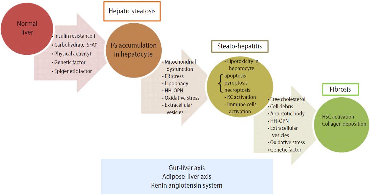
Hypertensive patients with biopsy-proven NAFLD on baseline RAS blockers had less advanced hepatic fibrosis [129]. Recently, a large retrospective study showed that angiotensin-converting enzyme inhibitors/angiotensin receptor blockers were associated with lower risk of hepatocellular carcinoma and cirrhotic complications in patients with NAFLD [130]. These data suggested a beneficial effect of RAS blockers in NAFLD. Transgenic hypertensive rats overexpressing the mouse renin gene with elevated levels of tissue angiotensin II developed hepatic steatosis, inflammation, and fibrosis [131]. The mice lacking the renin gene fed with high-fat diet had decreased liver fat [132]. Aliskiren, a direct renin inhibitor, reduced hepatic steatosis in high-fat diet-fed mice and fibrosis in mice fed with methionine-choline-deficient diet [133,134]. When renin or prorenin binds to the (pro)renin receptor (PRR), in addition to increasing the production and role of angiotensin (ANG II dependent pathway), it activates TGFβ, plasminogen activator inhibitor-1 (PAI-1), fibronectin, and collagen I independently from Ang II (ANG II independent pathway) [135-137]. Our group found that PRR contributed to liver fibrosis and HSC activation, and its down-regulation attenuated liver fibrosis through inactivation of the ERK/TGFβ1/Smad3 pathway [138]. These results indicate that renin and prorenin can directly activate renin (pro) receptor-related intracellular signaling pathways, including ERK, TGFβ, cyclooxygenease2, fibronectin, collagen I, and PAI-1 independently of angiotensin II to induce fibrosis. Moreover, Ren et al. [139] used N-acetylgalactosamine modified antisense oligonucleotides to suppress PRR expression in hepatocytes of high-fat diet-fed C57BL/6 mice, and found that PRR inhibition reduced acetyl-CoA carboxylase and pyruvate desorption hydrogenase protein expression. This change reprogrammed liver lipid metabolism, resulting in reduced lipid synthesis and increased fatty acid oxidation. As a result, liver PRR suppression attenuated dietinduced obesity and fatty liver [139]. The proposed pathogenesis that is involved from steatosis to fibrosis in patients with NAFLD is shown in Figure 1.

PROGRESSION OF NASH TO HCC

NASH is now the most common risk factor for HCC in the United States [140]. The potential pathways linking NASH to HCC include chronic inflammation of the liver [141], alternations in immune response, lipid metabolism and gut microbiome [142], and genetic factors. Enhanced IL-6 and TNF production during NAFLD cause hepatic inflammation and activation of the oncogenic transcription factor STAT3 [143]. ER stress contributes to NASH-driven hepatic tumorigenesis via TNFR1 [144]. The hepatic oxidative DNA damage was increased in patients with NASH who developed HCC [145]. The unconventional prefoldin RPB5 interactor-induced DNA damage in hepatocytes triggered inflammation via T helper 17 lymphocytes and interleukin 17A, contributing to NASH and HCC development [146]. Furthermore, NAFLD caused a selective loss of intrahepatic CD4(+) but not CD8(+) T lymphocytes, which led to accelerated hepatocarcinogenesis [147]. Neutrophil infiltration was characterized in NASH-HCC and can exist in both tumor promoting and suppressing states [148]. Fatty acid accumulation increased junctional protein associated with coronary artery disease, leading to the activation of Yes-associated protein 1 and tumor growth [149]. Dysregulated mammalian target of rapamycin stimulated sphingolipid and glycerophospholipid synthesis, leading to steatosis and HCC [150]. In NASH-driven HCC, metabolic reprogramming mediated by the downregulation of carnitine palmitoyltransferase 2 enables HCC cells to escape lipotoxicity and promotes hepatocarcinogenesis [151]. MicroRNA-21 can promote hepatic lipid accumulation and cancer progression by interacting with the sHbp1-p53-Srebp1c pathway [152]. The intestinal dysbiosis, gut permeability changes, and lipopolysaccharides translocation to the liver in NASH may increase secretion of the epiregulin growth factor, which triggers tumor hepatocyte proliferation [153]. Moreover, carriage of the PNPLA3 rs738409 C>G polymorphism is associated with a greater risk of NASH-HCC [154].

TREATMENT FOR NASH

Non-pharmacological therapy

Lifestyle modification

Lifestyle changes by eating less and exercising more to achieve weight loss remain the cornerstone of clinical care. Hypocaloric diet with a reduction of body weight decreased total body fat, visceral fat, and intrahepatic lipid content [155]. Some existing guidelines suggest restriction of energy by 1,200–1,500 kcal/day or a reduction of 500–1,000 kcal/day to achieve weight loss [1,156-158]. Weight reduction is beneficial for both non-obese (3–10%) [159] and obese patients (≥0%) [160,161]. Other dietary compositions that may be beneficial for NAFLD includes omega-3 PUFA and coffee. Omega-3 PUFA has been shown to increase insulin sensitivity [162] and ameliorate steatohepatitis in experimental studies [163,164]. One meta-analysis involving nine studies with 355 patients showed decreased liver fat in patients with PUFA treatment [165]. Coffee is not only associated with a reduced risk of NAFLD but also decreased risk of liver fibrosis among patients with NAFLD [166,167]. Regular exercise helps to enhance the effects of diet modifications. Physical activity with a target at least 150 min/week of moderate-intensity or 75–150 min/week of vigorous-intensity aerobic exercise is suggested [1,156-158]. Both aerobic and resistance exercises reduce the hepatic fat content [168,169]. In addition, the intensity of exercise may be more important than the duration or total volume [170]. In conclusion, lifestyle interventions to promote weight loss, which include both diet and exercise, are proven therapeutic strategies to improve fatty liver disease.

Surgery

Bariatric surgery provides sustained and durable weight loss and improving obesity-related diseases [171,172]. Currently, the most commonly performed bariatric procedures are laparoscopic sleeve gastrectomy (LSG) and Roux-en-Y gastric bypass. Two meta-analyses showed that bariatric surgery resulted in a biopsy-confirmed resolution of steatosis (56–66%), inflammation (45–50%), ballooning degeneration (49–76%), and fibrosis (25–40%), as well as reduction of NAFLD activity score (NAS) [173,174]. A higher rate of improvement in steatosis and hepatic fibrosis was observed in Asian countries compared to non-Asian countries [174]. In addition, bariatric surgery was associated with decreased progression of NAFLD to cirrhosis [175] and reduced risks of any cancer and obesity-related cancer in NAFLD patients with severe obesity, particularly in cirrhotic patients [176]. However, new or worsening cases of NAFLD were found in 12% of patients after bariatric surgery [173]. Bariatric surgery was associated with a significantly lower risk of incident major adverse liver outcomes (2.3% vs. 9.6% at 10 years) and major adverse cardiovascular events (8.5% vs. 15.7% at 10 years), as compared with non-surgical management [177].

Endoscopic therapy

Endoscopic bariatric therapies, including intragastric balloons (IGB), endoscopic sleeve gastroplasty (ESG), duodenal mucosal resurfacing (DMR), and duodenal-Jejunal bypass liner (DJBL), were recently introduced as less invasive modalities to treat obesity and metabolic comorbidities. In a meta-analysis, improvement in steatosis and NAS were seen in 79.2% and 83.5% of patients receiving IGB, respectively [178]. Improvement of fibrosis for 1.5 stage by MR elastography was seen in 50% of patients with NAFLD after IGB placement [179]. ESG reduced the body weight by up to 15% and improved hepatic steatosis and fibrosis at 2 years of follow-up in obese patients with NAFLD [180]. Studies for efficacy and safety of ESG (NCT03426111; NCT04653311) and the comparison of ESG vs. LSG (NCT04060368) in patients with NASH are ongoing. DMR has been shown to reduce alanine aminotransferase, aspartate aminotransferase, and fibrosis-4 scores in patients with diabetes mellitus [181]. Recently, an observational study of 32 obese patients with diabetes mellitus who underwent DJBL showed improved non-invasive markers of steatosis and NASH, but not fibrosis. The role of DJBL on NAFLD needs to be further evaluated [182].

Fecal microbiota transplantation

Some studies have suggested that fecal transplantation helps ameliorate steatohepatitis [183,184]. A randomized controlled trial (RCT) using allogenic fecal microbiota transplantation (FMT) from lean vegan donors for patients with NAFLD through duodenal infusion found that there was no significant improvement in NAS, steatosis, and fibrosis scores. However, they observed a trend of improving necro-inflammatory scores and beneficial changes in hepatic gene expression and plasma metabolites involved in inflammation and lipid metabolism following allogenic FMT [185]. Another RCT using allogenic FMT via endoscopic duodenal infusion in patients with NAFLD found that FMT did not improve insulin resistance and hepatic steatosis but reduced small intestinal permeability at 6 months of follow-up [186].

Pharmacological therapy

The pharmacological agents predominantly target the following four mechanisms: 1) hepatic fat accumulation; 2) oxidative stress, inflammation, and apoptosis; 3) gut-liver axis, including bile acids, gut microbiomes, and metabolic endotoxemia; and 4) hepatic fibrosis [187]. The agents targeting different pathways are described below, and those with promising results are summarized in Table 1.

Agents targeting hepatic fat accumulation

Pioglitazone, a PPAR γ agonist, improved hepatic steatosis, inflammation, and hepatocellular ballooning [188,189]. Similar effects were found in Asian NASH patients [190]. In the phase 3 RESOLVE-IT trial, Elafibranor, a dual PPAR α/δ agonist, failed to achieve NASH resolution [191]. Pemafibrate, a selective PPAR α modulator, did not decrease liver fat but caused a significant reduction in fibrosis for 6.2% of magnetic resonance elastography-based liver stiffness [192]. Lanifibranor, a pan-PPAR agonist, significantly decreased the steatosis-activity-fibrosis activity score for at least 2 points in 55% of the patients at 24 weeks [193].
GLP-1 agonists increase insulin secretion, inhibit glucagon secretion, delay gastric emptying, and decrease appetite. NASH resolution was observed in 39% of patients who received liraglutide for 48 weeks and in 59% of patients who received semaglutide for 72 weeks [194,195]. However, fibrosis improvement was insignificant in both studies.
Sodium-glucose cotransporter 2 (SGLT2) inhibitors increase the urinary excretion of glucose. A meta-analysis of 10 RCTs showed that SGLT2 inhibitors can reduce aminotransferases and hepatic fat [196].
FGF19 and FGF21 are endocrines that regulate energy homeostasis. Aldafermin, a FGF19 analogue, led to reductions of liver fat content and a trend toward fibrosis improvement [197]. Pegbelfermin and efruxifermin are long-acting, recombinant analogues of human FGF21, and both have shown effects of reducing liver fat [198,199].
Two phase IIa trials investigated the effects of acetyl-coenzyme A carboxylase (ACC) inhibitor monotherapy (PF-05221304) and combination with a diacylglycerol O-acyltransferase 2 (DGAT2) inhibitor (PF-06865571). Both PF-05221304 monotherapy and co-administration with PF-06865571 reduced liver fat content [200].
Stearoyl-coenzyme A desaturase 1 (SCD-1) is a key enzyme that catalyzes the biosynthesis of monounsaturated fatty acids. A phase IIb trial (ARREST trial) showed that aramchol (a liver-targeted SCD-1 inhibitor) 600 mg did not cause a significant reduction in liver fat content. Nevertheless, the observed change in liver histology and biochemical improvement suggests a potential role of aramchol in treating NASH and fibrosis [201].
Thyroid hormone receptor-β (THR-β) is predominantly expressed in the hepatocytes. Resmetirom, a selective THR-β agonist, significantly reduced more than 30% of hepatic fat after 12 and 36 weeks of treatment in patients with NASH in phase II trial [202].

Agents targeting oxidative stress, inflammation, and apoptosis

Vitamin E, an antioxidative agent, demonstrated benefits on hepatic decompensation and transplant-free survival in patient with NASH [203]. The PIVENS study, which compared the effects of vitamin E, pioglitazone, and placebo in NASH patients without diabetes, showed that vitamin E (800 international units/day), but not pioglitazone, significantly improved NASH [204].
Apoptosis signaling kinase 1 (ASK1) promotes apoptosis, inflammation, and fibrosis in the liver. However, selonsertib, an ASK1 inhibitor, failed to improve fibrosis in NASH patients with bridging fibrosis or compensated cirrhosis [205].
Berberine ursodeoxycholate is an ionic salt of berberine and ursodeoxycholic acid. It reduced 4.8% of liver fat and improved glycemic control as well as liver enzymes in patients with NASH and diabetes [206].

Agents targeting gut-liver axis

In a phase IIb study, obeticholic acid (OCA), a FXR agonist, improved liver histology in 21% of NASH patients [207]. In patients with NASH and diabetes, OCA demonstrated the effects of increasing insulin sensitivity and reducing markers of liver inflammation as well as fibrosis [208]. In the interim analysis of a phase III trial, both 10-mg and 25-mg doses of OCA improved fibrosis (18% and 23%, respectively), but the NASH resolution endpoint was not met [209]. This study is ongoing to assess the clinical outcomes.

Agents targeting liver fibrosis

Caspase is a protease that is associated with apoptosis and inflammation in the liver. However, emricasan, a pan-caspase inhibitor, did not improve fibrosis or resolution of NASH [210]. Besides, for patients with NASH-related cirrhosis and severe portal hypertension, emricasan did not improve hepatic venous pressure gradient (HVPG) or liver-related outcomes [211].
In a phase IIb CENTAUR trial, a 2-year study, cenicriviroc, a dual C-C chemokine receptor types 2 and 5 antagonist, achieved ≥1-stage of fibrosis improvement without worsening of NASH after 1 year of treatment compared to placebo (20% vs. 10%) [212]. And a great proportion (60%) of the patients who achieved fibrosis response at the first year maintained fibrosis reduction at the second year [213]. The long-term impact of cenicriviroc on fibrosis needs to be further investigated.
Belapectin, a galectin-3 inhibitor, did not significantly reduce HVPG or fibrosis in patients with NASH, cirrhosis, and portal hypertension; however, in a subgroup of patients without esophageal varices, belapectin reduced HVPG as well as the development of esophageal varices [214].
Information about the ongoing phase III clinical trials of promising drugs on phase II studies are listed in Table 2.

Combination therapy

NAFLD is a multifactorial disease, and combining therapies with different targets may have synergistic effects [215]. Cilofexor (FXR agonist) plus firsocostat (ACC inhibitor) led to improvements in NASH activity compared to placebo, or single agent in patients with bridging fibrosis and cirrhosis [216]. Semaglutide with firsocostat and/or cilofexor showed greater improvements in liver steatosis and liver biochemistry compared to semaglutide alone [217]. Combining ACC inhibitors and DGAT2 inhibitors reduced liver fat content and mitigated the side effect of elevated serum TGs [200].

PERSPECTIVES

As understanding of mechanisms of NASH and its fibrosis increases, more therapies will be introduced and tested in clinical trials. The pathogenesis of NASH and fibrosis is complex; therefore, it would be difficult to treat the disease using just one therapy. Combination therapy is the focus in the future development of treatment. Furthermore, better care of extra-hepatic complications of NASH, novel biomarkers for diagnosis, risk stratification and treatment responses, and more clinical trials in Asian groups should also be well researched and developed.

ACKNOWLEDGMENTS

This study was supported in part by the grants from the Ministry of Science and Technology (110-2628-B-075-016; 111-2314-B-075-051-MY3) Taiwan.

FOOTNOTES

Authors’ contribution
KC Lee and PS Wu drafted the manuscript and HC Lin revised the manuscript. All the authors read and approved the final version.
Conflicts of Interest
The authors have no conflicts to disclose.

Figure 1.
Progression of hepatic steatosis to inflammation and fibrosis in liver. Both metabolic and genetic factors contribute to the formation of hepatic steatosis. Fat accumulation in hepatocytes leads to organelles dysfunction and lipotoxicity. Then, oxidative stress species or signaling molecules are transmitted through extracellular vesicles or diffusion, activating other parenchymal and non-parenchymal cells, which subsequently causes inflammatory cascades, steatohepatitis, and liver fibrosis. On the other hand, gut-derived bacterial end-products, metabolites, gut hormones, adipose tissue-derived cytokines or adipokines, and renin-angiotensin-system all contribute to the progression from steatosis to inflammation and fibrosis. SFA, saturated fatty acid; TG, triglyceride; ER, endoplastic reticulum; HH-OPN, Hedgehog-osteopontin; KC, Kupffer cell; HSC, hepatic stellate cell.

cmh-2022-0237f1.jpg
Table 1.
Promising pharmacological therapies for NAFLD or NASH
Type of drug Mechanism of action Drug name Study design Study outcome Reference
PPAR agonist PPAR-γ: ↑ insulin sensitivity modulates adipose tissue distribution Pioglitazone (PPAR-γ agonist) RCT; NASH, prediabetes/DM ↓ ALT/AST [188]
Pioglitazone vs. placebo ↓ Steatosis, ballooning necrosis, and inflammation [189]
PPAR-α: ↑ fatty acid β-oxidation Fibrosis not improved
PPAR-δ: anti-inflammatory RCT; NASH ↓ ALT/GGT
Pioglitazone vs. placebo Histology improvement, including liver injury and fibrosis
Pemafibrate (SPPARMα) Phase II RCT; NAFLD and ↑ ALT ↓ ALT [192]
↓ Liver stiffness
Pemafibrate vs. placebo No significant change of liver fat
Lanifibranor (Pan-PPAR agonist) Phase IIb RCT; NASH (SAF-A ≥3) ↓ SAF-A score ≥2 points in lanifibranor 1,200 mg group [193]
Lanifibranor vs. placebo
GLP-1 agonist ↑ Insulin secretion Liraglutide Phase II RCT; NASH, obesity ↑ Resolution of NASH without worsening of fibrosis [194]
↓ Glucagon secretion Liraglutide vs. placebo No difference in fibrosis improvement
↓ Gastric emptying Semaglutide RCT, phase II; NASH (F1–F3 fibrosis) ↑ Resolution of NASH without worsening of fibrosis in semaglutide 0.4 mg group [195]
↓ Appetite Semaglutide vs. placebo No difference in fibrosis improvement
SGLT2 inhibitor ↑ Urinary excretion of glucose SGLT2 inhibitors Meta-analysis of 10 RCTs; NAFLD, DM ↓ ALT/AST [196]
SGLT2 inhibitor vs. other antidiabetic drugs ↓ Liver fat content, visceral fat, and subcutaneous fat areas
FGF-19 analogue ↓ Bile acids synthesis Aldafermin Phase II RCT; NASH (NAS ≥4, F2–F3 fibrosis, and liver fat content ≥8%) ↓ ALT/AST [197]
↓ Hepatic gluconeogenesis ↓ Liver fat fraction on MRI-PDFF
↓ DNL Aldafermin vs. placebo
↑ Fatty acid oxidation
FGF-21 analogue ↓ Hepatic gluconeogenesis Pegbelfermin Phase IIa RCT; NASH (F1–F3 fibrosis, and liver fat content ≥10%), obesity ↓ Liver fat fraction on MRI-PDFF [198]
↑ Insulin sensitivity
↑ Energy expenditure Pegbelfermin vs. placebo
↑ Mitochondria beta-oxidation in hepatocytes Efruxifermin Phase IIa RCT; NASH ↓ Liver fat fraction on MRI-PDFF [199]
Efruxifermin vs. placebo
Acetyl-CoA carboxylase inhibitor ↓ DNL PF-05221304 2 phase IIa RCTs; NAFLD/NASH PF-05221304 monotherapy: [200]
↑ Fatty acid oxidation PF-05221304 monotherapy vs. placebo ↓Liver fat on MRI-PDFF, but ↑ TG
Diacylglycerol acyltransferase 2 inhibitor ↓ Synthesis of fatty acids into TGs PF-06865571 PF-05221304 and PF-06865571 co-administration vs. placebo Co-administration therapy: ↓ liver fat and mitigated ACC inhibitormediated effect on TG
Stearoyl-CoA desaturase 1 inhibitor ↓ DNL Aramchol Phase IIb RCT; NASH ↓ Liver fat content in aramchol 600 mg group, but not significant [201]
Aramchol vs. placebo
Selective thyroid hormone receptor-β agonist ↓ LDL, cholesterol, and TG Resmetirom Phase II RCT; NASH (F1–F3 fibrosis, and liver fat content ≥10%) ↓ Liver fat on MRI-PDFF [202]
↑ Fatty acid oxidation Resmetirom vs. placebo
Anti-oxidant Anti-oxidative stress Vitamin E RCT; NASH, no DM ↓ ALT/AST for both vitamin E and pioglitazone groups [204]
Vitamin E vs. pioglitazone vs. placebo NASH improvement in vitamin E group, but not in pioglitazone group
Fibrosis not improved in vitamin E and pioglitazone groups
Bile acid analogue Anti-inflammation Berberine ursodeoxycholate Phase II RCT; NAFLD, DM ↓ Liver fat content on MRI-PDFF [206]
Berberine ursodeoxycholate vs. placebo
↓ DNL Obeticholic acid (FXR agonist) Phase III RCT; NASH (NAS ≥4, F2–F3 fibrosis or F1 with ≥1 accompanying comorbidity) ↓ Fibrosis [209]
↑ Fatty acid β-oxidation No significant resolution of NASH
↑ Cholesterol excretion Obeticholic acid vs. placebo

NAFLD, non-alcoholic fatty liver disease; NASH, non-alcoholic steatohepatitis; PPAR, proliferator-activated receptor; SPPARMα, selective peroxisome proliferator-activated receptor α modulator; RCT, randomized controlled trial; ALT, alanine aminotransferase; SAF-A, steatosis-activity-fibrosis activity; GLP-1, glucagon-like peptide-1; SGLT2, sodium-glucose cotransporter 2; DM, diabetes mellitus; AST, aspartate aminotransferase; FGF, fibroblast growth factor; DNL, de novo lipogenesis; NAS, NAFLD activity score; MRI, magnetic resonance imaging; PDFF, proton density fat fraction; TG, triglyceride; ACC, acetyl-coenzyme A carboxylase; LDL, low density lipoprotein; FXR, farnesoid X receptor.

Table 2.
Ongoing phase III clinical trials of pharmacological agents in patients with NAFLD or NASH
Agent Mechanism Patient Outcome Status ClinicalTrials.gov identifier
Lanifibranor Pan-PPAR agonist NASH with stage 2–3 fibrosis without cirrhosis NASH resolution; fibrosis improvement; liver-related events Recruiting NCT04849728
Semaglutide GLP-1 agonist NASH with stage 2–3 fibrosis NASH resolution; fibrosis improvement; liver-related events Recruiting NCT04822181
Dapagliflozin SGLT2 inhibitor NASH and type 2 DM without cirrhosis Histology improvement Recruiting NCT03723252
Resmetirom Selective thyroid hormone receptor-β agonist NASH without cirrhosis NASH resolution; liver-related events Recruiting NCT03900429
Aramchol Stearoyl-CoA desaturase 1 inhibitor NASH with stage 2–3 fibrosis without cirrhosis; type 2 DM or prediabetes NASH resolution; fibrosis improvement Recruiting NCT04104321
Belapectin Galectin-3 inhibitor NASH cirrhosis without esophageal or gastric varices Newly developed esophageal varices Recruiting NCT04365868
Oltipraz Liver X receptor alpha-inhibitor NAFLD without cirrhosis Liver fat Recruiting NCT04142749

NAFLD, non-alcoholic fatty liver disease; NASH, non-alcoholic steatohepatitis; PPAR, proliferator-activated receptor; GLP-1, glucagon-like peptide-1; SGLT2, sodium-glucose cotransporter 2; DM, diabetes mellitus.

Abbreviations

ACC
acetyl-coenzyme A carboxylase
AMPK
AMP-activated protein kinase
ANG
angiotensin
ASK1
apoptosis signaling kinase 1
CHOP
C/EBP homologous protein
DGAT2
diacylglycerol O-acyltransferase 2
DJBL
duodenal-Jejunal bypass liner
DMR
duodenal mucosal resurfacing
DNL
de novo lipogenesis
ER
endoplasmic reticulum
ESG
endoscopic sleeve gastroplasty
FFA
free fatty acid
FGF
fibroblast growth factor
FMT
fecal microbiota transplantation
FXR
farnesoid X receptor
GCKR
glucokinase regulatory protein
GLP-1
glucagon-like peptide-1
HCC
hepatocellular carcinoma
Hh
hedgehog
HSC
hepatic stellate cell
HSD17B13
hydroxysteroid 17- beta dehydrogenase 13
HVPG
hepatic venous pressure gradient
IGB
intragastric balloons
IL
interleukin
IRE1
inositol-requiring enzyme 1
IRGM
immunity-related GTPase M
KC
Kupffer cell
LSG
laparoscopic sleeve gastrectomy
MBOAT7
membrane-bound O-acyltransferase domain-containing protein 7
miRNA
microRNA
NAFLD
non-alcoholic fatty liver disease
NAS
NAFLD activity score
NASH
non-alcoholic steatohepatitis
NEFA
non-esterified fatty acid
OCA
obeticholic acid
PAI-1
plasminogen activator inhibitor-1
PERK
protein kinase R (double-stranded RNA-activated protein kinase)-like ER kinases
PNPLA3
patatin-like phospholipase domaincontaining 3
PPAR
proliferator-activated receptor
PRR
(pro)renin receptor
PUFA
polyunsaturated fatty acid
RAS
renin-angiotensin system
RCT
randomized controlled trial
SCD-1
stearoyl-coenzyme A desaturase 1
SCFA
short-chain fatty acid
SFA
saturated fatty acid
SGLT2
sodium-glucose cotransporter 2
SNP
single nucleotide polymorphism
SREBP1c
sterol receptor binding protein 1-c
TG
triglyceride
TGF
transforming growth factor
THR-β
thyroid hormone receptor-β
TLR
toll-like receptor
TM6SF2
transmembrane 6 superfamily 2
TNF
tumor necrosis factor
TNFR1
TNF receptor 1
UPR
unfolded protein response
VLDL
very-low-density lipoprotein
XBP-1
X-box-binding protein 1
ω6
omega-6

REFERENCES

1. Chalasani N, Younossi Z, Lavine JE, Charlton M, Cusi K, Rinella M, et al. The diagnosis and management of nonalcoholic fatty liver disease: practice guidance from the American Association for the Study of Liver Diseases. Hepatology 2018;67:328-357.
crossref pmid pdf
2. Browning JD, Horton JD. Molecular mediators of hepatic steatosis and liver injury. J Clin Invest 2004;114:147-152.
crossref pmid pmc
3. Lemoinne S, Friedman SL. New and emerging anti-fibrotic therapeutics entering or already in clinical trials in chronic liver diseases. Curr Opin Pharmacol 2019;49:60-70.
crossref pmid
4. Westerbacka J, Lammi K, Häkkinen AM, Rissanen A, Salminen I, Aro A, et al. Dietary fat content modifies liver fat in overweight nondiabetic subjects. J Clin Endocrinol Metab 2005;90:2804-2809.
crossref pmid
5. van der Meer RW, Hammer S, Lamb HJ, Frölich M, Diamant M, Rijzewijk LJ, et al. Effects of short-term high-fat, high-energy diet on hepatic and myocardial triglyceride content in healthy men. J Clin Endocrinol Metab 2008;93:2702-2708.
crossref pmid pdf
6. Lindeboom L, Nabuurs CI, Hesselink MK, Wildberger JE, Schrauwen P, Schrauwen-Hinderling VB. Proton magnetic resonance spectroscopy reveals increased hepatic lipid content after a single high-fat meal with no additional modulation by added protein. Am J Clin Nutr 2015;101:65-71.
crossref pmid
7. Hernández EA, Kahl S, Seelig A, Begovatz P, Irmler M, Kupriyanova Y, et al. Acute dietary fat intake initiates alterations in energy metabolism and insulin resistance. J Clin Invest 2017;127:695-708.
crossref pmid pmc
8. Kaikkonen JE, Würtz P, Suomela E, Lehtovirta M, Kangas AJ, Jula A, et al. Metabolic profiling of fatty liver in young and middle-aged adults: cross-sectional and prospective analyses of the young finns study. Hepatology 2017;65:491-500.
crossref pmid pdf
9. Rosqvist F, Kullberg J, Ståhlman M, Cedernaes J, Heurling K, Johansson HE, et al. Overeating saturated fat promotes fatty liver and ceramides compared with polyunsaturated fat: a randomized trial. J Clini Endocrinol Metab 2019;104:6207-6219.
crossref pdf
10. Puri P, Baillie RA, Wiest MM, Mirshahi F, Choudhury J, Cheung O, et al. A lipidomic analysis of nonalcoholic fatty liver disease. Hepatology 2007;46:1081-1090.
crossref pmid
11. Gerber L, Otgonsuren M, Mishra A, Escheik C, Birerdinc A, Stepanova M, et al. Non-alcoholic fatty liver disease (NAFLD) is associated with low level of physical activity: a populationbased study. Aliment Pharmacol Ther 2012;36:772-781.
crossref pmid
12. Ryu S, Chang Y, Jung HS, Yun KE, Kwon MJ, Choi Y, et al. Relationship of sitting time and physical activity with non-alcoholic fatty liver disease. J Hepatology 2015;63:1229-1237.
crossref
13. Marchesini G, Brizi M, Bianchi G, Tomassetti S, Bugianesi E, Lenzi M, et al. Nonalcoholic fatty liver disease: a feature of the metabolic syndrome. Diabetes 2001;50:1844-1850.
pmid
14. Bugianesi E, Gastaldelli A, Vanni E, Gambino R, Cassader M, Baldi S, et al. Insulin resistance in non-diabetic patients with non-alcoholic fatty liver disease: sites and mechanisms. Diabetologia 2005;48:634-642.
crossref pmid pdf
15. Donnelly KL, Smith CI, Schwarzenberg SJ, Jessurun J, Boldt MD, Parks EJ. Sources of fatty acids stored in liver and secreted via lipoproteins in patients with nonalcoholic fatty liver disease. J Clin Invest 2005;115:1343-1351.
crossref pmid pmc
16. Utzschneider KM, Kahn SE. Review: the role of insulin resistance in nonalcoholic fatty liver disease. J Clin Endocrinol Metab 2006;91:4753-4761.
pmid
17. Shimano H, Horton JD, Shimomura I, Hammer RE, Brown MS, Goldstein JL. Isoform 1c of sterol regulatory element binding protein is less active than isoform 1a in livers of transgenic mice and in cultured cells. J Clin Invest 1997;99:846-854.
crossref pmid pmc
18. Tamura S, Shimomura I. Contribution of adipose tissue and de novo lipogenesis to nonalcoholic fatty liver disease. J Clin Invest 2005;115:1139-1142.
crossref pmid pmc
19. Wolfrum C, Asilmaz E, Luca E, Friedman JM, Stoffel M. Foxa2 regulates lipid metabolism and ketogenesis in the liver during fasting and in diabetes. Nature 2004;432:1027-1032.
crossref pmid pdf
20. Romeo S, Kozlitina J, Xing C, Pertsemlidis A, Cox D, Pennacchio LA, et al. Genetic variation in PNPLA3 confers susceptibility to nonalcoholic fatty liver disease. Nat Genet 2008;40:1461-1465.
crossref pmid pmc pdf
21. Speliotes EK, Yerges-Armstrong LM, Wu J, Hernaez R, Kim LJ, Palmer CD, et al. Genome-wide association analysis identifies variants associated with nonalcoholic fatty liver disease that have distinct effects on metabolic traits. PLoS Genet 2011;7:e1001324.
crossref pmid pmc
22. Chambers JC, Zhang W, Sehmi J, Li X, Wass MN, Van der Harst P, et al. Genome-wide association study identifies loci influencing concentrations of liver enzymes in plasma. Nat Genet 2011;43:1131-1138.
pmid pmc
23. Beer NL, Tribble ND, McCulloch LJ, Roos C, Johnson PR, OrhoMelander M, et al. The P446L variant in GCKR associated with fasting plasma glucose and triglyceride levels exerts its effect through increased glucokinase activity in liver. Hum Mol Genet 2009;18:4081-4088.
crossref pmid pmc
24. Kozlitina J, Smagris E, Stender S, Nordestgaard BG, Zhou HH, Tybjaerg-Hansen A, et al. Exome-wide association study identifies a TM6SF2 variant that confers susceptibility to nonalcoholic fatty liver disease. Nat Genet 2014;46:352-356.
crossref pmid pmc pdf
25. Mahdessian H, Taxiarchis A, Popov S, Silveira A, FrancoCereceda A, Hamsten A, et al. TM6SF2 is a regulator of liver fat metabolism influencing triglyceride secretion and hepatic lipid droplet content. Proc Natl Acad Sci U S A 2014;111:8913-8918.
crossref pmid pmc
26. Lin YC, Chang PF, Lin HF, Liu K, Chang MH, Ni YH. Variants in the autophagy-related gene IRGM confer susceptibility to non-alcoholic fatty liver disease by modulating lipophagy. J Hepatol 2016;65:1209-1216.
crossref pmid
27. Bellini G, Miraglia Del Giudice E, Nobili V, Rossi F. The IRGM rs10065172 variant increases the risk for steatosis but not for liver damage progression in Italian obese children. J Hepatol 2017;67:653-655.
crossref pmid
28. Luukkonen PK, Qadri S, Ahlholm N, Porthan K, Mannisto V, Sammalkorpi H, et al. Distinct contributions of metabolic dysfunction and genetic risk factors in the pathogenesis of nonalcoholic fatty liver disease. J Hepatol 2022;76:526-535.
crossref pmid pmc
29. Ma J, Nano J, Ding J, Zheng Y, Hennein R, Liu C, et al. A peripheral blood DNA methylation signature of hepatic fat reveals a potential causal pathway for nonalcoholic fatty liver disease. Diabetes 2019;68:1073-1083.
crossref pmid pmc pdf
30. Pirola CJ, Fernandez Gianotti T, Castano GO, Mallardi P, San Martino J, Mora Gonzalez Lopez Ledesma M, et al. Circulating microRNA signature in non-alcoholic fatty liver disease: from serum non-coding RNAs to liver histology and disease pathogenesis. Gut 2015;64:800-812.
crossref pmid
31. Castano C, Kalko S, Novials A, Parrizas M. Obesity-associated exosomal miRNAs modulate glucose and lipid metabolism in mice. Proc Natl Acad Sci U S A 2018;115:12158-12163.
crossref pmid pmc
32. Esau C, Davis S, Murray SF, Yu XX, Pandey SK, Pear M, et al. miR122 regulation of lipid metabolism revealed by in vivo antisense targeting. Cell Metab 2006;3:87-98.
crossref pmid
33. Latorre J, Moreno-Navarrete JM, Mercader JM, Sabater M, Rovira O, Girones J, et al. Decreased lipid metabolism but increased FA biosynthesis are coupled with changes in liver microRNAs in obese subjects with NAFLD. Int J Obes (Lond) 2017;41:620-630.
crossref pmid pdf
34. Hsu SH, Wang B, Kota J, Yu J, Costinean S, Kutay H, et al. Essential metabolic, anti-inflammatory, and anti-tumorigenic functions of miR-122 in liver. J Clin Invest 2012;122:2871-2883.
crossref pmid pmc
35. Kim YS, Nam HJ, Han CY, Joo MS, Jang K, Jun DW, et al. Liver X receptor alpha activation inhibits autophagy and lipophagy in hepatocytes by dysregulating autophagy-related 4B cysteine peptidase and Rab-8B, reducing mitochondrial fuel oxidation. Hepatology 2021;73:1307-1326.
crossref pmid pdf
36. Wang M, Kaufman RJ. Protein misfolding in the endoplasmic reticulum as a conduit to human disease. Nature 2016;529:326-335.
crossref pmid pdf
37. Lebeaupin C, Vallee D, Hazari Y, Hetz C, Chevet E, Bailly-Maitre B. Endoplasmic reticulum stress signalling and the pathogenesis of non-alcoholic fatty liver disease. J Hepatol 2018;69:927-947.
crossref
38. Puri P, Mirshahi F, Cheung O, Natarajan R, Maher JW, Kellum JM, et al. Activation and dysregulation of the unfolded protein response in nonalcoholic fatty liver disease. Gastroenterology 2008;134:568-576.
crossref pmid
39. Akazawa Y, Cazanave S, Mott JL, Elmi N, Bronk SF, Kohno S, et al. Palmitoleate attenuates palmitate-induced Bim and PUMA up-regulation and hepatocyte lipoapoptosis. J Hepatol 2010;52:586-593.
crossref pmid pmc
40. Volmer R, van der Ploeg K, Ron D. Membrane lipid saturation activates endoplasmic reticulum unfolded protein response transducers through their transmembrane domains. Proc Natl Acad Sci U S A 2013;110:4628-4633.
crossref pmid pmc
41. Latif MU, Schmidt GE, Mercan S, Rahman R, Gibhardt CS, Stejerean-Todoran I, et al. NFATc1 signaling drives chronic ER stress responses to promote NAFLD progression. Gut 2022;71:2561-2573.
crossref pmid
42. Sunny NE, Parks EJ, Browning JD, Burgess SC. Excessive hepatic mitochondrial TCA cycle and gluconeogenesis in humans with nonalcoholic fatty liver disease. Cell Metab 2011;14:804-810.
crossref pmid pmc
43. Koliaki C, Szendroedi J, Kaul K, Jelenik T, Nowotny P, Jankowiak F, et al. Adaptation of hepatic mitochondrial function in humans with non-alcoholic fatty liver is lost in steatohepatitis. Cell Metab 2015;21:739-746.
crossref pmid
44. Perez-Carreras M, Del Hoyo P, Martin MA, Rubio JC, Martin A, Castellano G, et al. Defective hepatic mitochondrial respiratory chain in patients with nonalcoholic steatohepatitis. Hepatology 2003;38:999-1007.
crossref pmid
45. Satapati S, Kucejova B, Duarte JA, Fletcher JA, Reynolds L, Sunny NE, et al. Mitochondrial metabolism mediates oxidative stress and inflammation in fatty liver. J Clin Invest 2015;125:4447-4462.
crossref pmid pmc
46. Martel C, Allouche M, Esposti DD, Fanelli E, Boursier C, Henry C, et al. Glycogen synthase kinase 3-mediated voltage-dependent anion channel phosphorylation controls outer mitochondrial membrane permeability during lipid accumulation. Hepatology 2013;57:93-102.
crossref pmid
47. Li Z, Berk M, McIntyre TM, Gores GJ, Feldstein AE. The lysosomal-mitochondrial axis in free fatty acid-induced hepatic lipotoxicity. Hepatology 2008;47:1495-1503.
crossref pmid
48. Meex RCR, Blaak EE. Mitochondrial dysfunction is a key pathway that links saturated fat intake to the development and progression of NAFLD. Mol Nutr Food Res 2021;65:e1900942.
crossref pmid pdf
49. Peng KY, Watt MJ, Rensen S, Greve JW, Huynh K, Jayawardana KS, et al. Mitochondrial dysfunction-related lipid changes occur in nonalcoholic fatty liver disease progression. J Lipid Res 2018;59:1977-1986.
crossref pmid pmc
50. Chocian G, Chabowski A, Zendzian-Piotrowska M, Harasim E, Lukaszuk B, Gorski J. High fat diet induces ceramide and sphingomyelin formation in rat’s liver nuclei. Mol Cell Biochem 2010;340:125-131.
crossref pmid pdf
51. Chavez JA, Summers SA. A ceramide-centric view of insulin resistance. Cell Metab 2012;15:585-594.
crossref pmid
52. Arora AS, Jones BJ, Patel TC, Bronk SF, Gores GJ. Ceramide induces hepatocyte cell death through disruption of mitochondrial function in the rat. Hepatology 1997;25:958-963.
crossref pmid
53. Tovoli F, Napoli L, Negrini G, D’Addato S, Tozzi G, D’Amico J, et al. A relative deficiency of lysosomal acid lypase activity characterizes non-alcoholic fatty liver disease. Int J mol Sci 2017;18:1134.
crossref pmid pmc
54. Baratta F, Pastori D, Del Ben M, Polimeni L, Labbadia G, Di Santo S, et al. Reduced lysosomal acid lipase activity in adult patients with non-alcoholic fatty liver disease. EBioMedicine 2015;2:750-754.
crossref pmid pmc
55. Wang X, Zhang X, Chu ESH, Chen X, Kang W, Wu F, et al. Defective lysosomal clearance of autophagosomes and its clinical implications in nonalcoholic steatohepatitis. FASEB J 2018;32:37-51.
crossref pmid pdf
56. Feldstein AE, Werneburg NW, Canbay A, Guicciardi ME, Bronk SF, Rydzewski R, et al. Free fatty acids promote hepatic lipotoxicity by stimulating TNF-alpha expression via a lysosomal pathway. Hepatology 2004;40:185-194.
crossref pmid
57. Hirsova P, Ibrabim SH, Gores GJ, Malhi H. Lipotoxic lethal and sublethal stress signaling in hepatocytes: relevance to NASH pathogenesis. J Lipid Res 2016;57:1758-1770.
crossref pmid pmc
58. Singh R, Kaushik S, Wang Y, Xiang Y, Novak I, Komatsu M, et al. Autophagy regulates lipid metabolism. Nature 2009;458:1131-1135.
crossref pmid pmc pdf
59. Li S, Dou X, Ning H, Song Q, Wei W, Zhang X, et al. Sirtuin 3 acts as a negative regulator of autophagy dictating hepatocyte susceptibility to lipotoxicity. Hepatology 2017;66:936-952.
crossref pmid pdf
60. Geng Y, Faber KN, de Meijer VE, Blokzijl H, Moshage H. How does hepatic lipid accumulation lead to lipotoxicity in nonalcoholic fatty liver disease? Hepatol Int 2021;15:21-35.
crossref pmid pdf
61. Cao L, Quan XB, Zeng WJ, Yang XO, Wang MJ. Mechanism of hepatocyte apoptosis. J Cell Death 2016;9:19-29.
crossref pmid pmc pdf
62. Rada P, Gonzalez-Rodriguez A, Garcia-Monzon C, Valverde AM. Understanding lipotoxicity in NAFLD pathogenesis: is CD36 a key driver? Cell Death Dis 2020;11:802.
crossref pmid pmc pdf
63. Sanyal AJ, Van Natta ML, Clark J, Neuschwander-Tetri BA, Diehl A, Dasarathy S, et al. Prospective study of outcomes in adults with nonalcoholic fatty liver disease. N Engl J Med 2021;385:1559-1569.
crossref pmid pmc
64. Kim Y, Chang Y, Cho YK, Ahn J, Shin H, Ryu S. Obesity and weight gain are associated with progression of fibrosis in patients with nonalcoholic fatty liver disease. Clin Gastroenterol Hepatol 2019;17:543-550 e2.
crossref pmid
65. Singh S, Allen AM, Wang Z, Prokop LJ, Murad MH, Loomba R. Fibrosis progression in nonalcoholic fatty liver vs nonalcoholic steatohepatitis: a systematic review and meta-analysis of paired-biopsy studies. Clin Gastroenterol Hepatol 2015;13:643-654 e1-e9.
crossref pmid
66. Argo CK, Northup PG, Al-Osaimi AM, Caldwell SH. Systematic review of risk factors for fibrosis progression in non-alcoholic steatohepatitis. J Hepatol 2009;51:371-379.
crossref pmid
67. Parthasarathy G, Revelo X, Malhi H. Pathogenesis of nonalcoholic steatohepatitis: an overview. Hepatol Commun 2020;4:478-492.
crossref pmid pmc pdf
68. Marra F, Svegliati-Baroni G. Lipotoxicity and the gut-liver axis in NASH pathogenesis. J Hepatol 2018;68:280-295.
crossref pmid
69. Hirsova P, Ibrahim SH, Krishnan A, Verma VK, Bronk SF, Werneburg NW, et al. Lipid-induced signaling causes release of inflammatory extracellular vesicles from hepatocytes. Gastroenterology 2016;150:956-967.
crossref pmid
70. Canbay A, Taimr P, Torok N, Higuchi H, Friedman S, Gores GJ. Apoptotic body engulfment by a human stellate cell line is profibrogenic. Lab Invest 2003;83:655-663.
crossref pmid
71. Kwon H, Song K, Han C, Chen W, Wang Y, Dash S, et al. Inhibition of hedgehog signaling ameliorates hepatic inflammation in mice with nonalcoholic fatty liver disease. Hepatology 2016;63:1155-1169.
crossref pmid pdf
72. Syn WK, Jung Y, Omenetti A, Abdelmalek M, Guy CD, Yang L, et al. Hedgehog-mediated epithelial-to-mesenchymal transition and fibrogenic repair in nonalcoholic fatty liver disease. Gastroenterology 2009;137:1478-1488 e8.
crossref pmid
73. Syn WK, Choi SS, Liaskou E, Karaca GF, Agboola KM, Oo YH, et al. Osteopontin is induced by hedgehog pathway activation and promotes fibrosis progression in nonalcoholic steatohepatitis. Hepatology 2011;53:106-115.
crossref pmid
74. Syn WK, Agboola KM, Swiderska M, Michelotti GA, Liaskou E, Pang H, et al. NKT-associated hedgehog and osteopontin drive fibrogenesis in non-alcoholic fatty liver disease. Gut 2012;61:1323-1329.
crossref pmid
75. Shi H, Kokoeva MV, Inouye K, Tzameli I, Yin H, Flier JS. TLR4 links innate immunity and fatty acid-induced insulin resistance. J Clin Invest 2006;116:3015-3025.
crossref pmid pmc
76. Hetherington AM, Sawyez CG, Zilberman E, Stoianov AM, Robson DL, Borradaile NM. Differential lipotoxic effects of palmitate and oleate in activated human hepatic stellate cells and epithelial hepatoma cells. Cell Physiol Biochem 2016;39:1648-1662.
crossref pmid pdf
77. Tomita K, Teratani T, Suzuki T, Shimizu M, Sato H, Narimatsu K, et al. Free cholesterol accumulation in hepatic stellate cells: mechanism of liver fibrosis aggravation in nonalcoholic steatohepatitis in mice. Hepatology 2014;59:154-169.
crossref pmid
78. Paradis V, Perlemuter G, Bonvoust F, Dargere D, Parfait B, Vidaud M, et al. High glucose and hyperinsulinemia stimulate connective tissue growth factor expression: a potential mechanism involved in progression to fibrosis in nonalcoholic steatohepatitis. Hepatology 2001;34:738-744.
crossref pmid
79. Svegliati-Baroni G, Ridolfi F, Di Sario A, Casini A, Marucci L, Gaggiotti G, et al. Insulin and insulin-like growth factor-1 stimulate proliferation and type I collagen accumulation by human hepatic stellate cells: differential effects on signal transduction pathways. Hepatology 1999;29:1743-1751.
crossref pmid
80. Cai CX, Buddha H, Castelino-Prabhu S, Zhang Z, Britton RS, Bacon BR, et al. Activation of insulin-PI3K/akt-p70S6K pathway in hepatic stellate cells contributes to fibrosis in nonalcoholic steatohepatitis. Dig Dis Sci 2017;62:968-978.
crossref pmid pdf
81. Pirazzi C, Valenti L, Motta BM, Pingitore P, Hedfalk K, Mancina RM, et al. PNPLA3 has retinyl-palmitate lipase activity in human hepatic stellate cells. Hum Mol Genet 2014;23:4077-4085.
crossref pmid pmc
82. Mancina RM, Dongiovanni P, Petta S, Pingitore P, Meroni M, Rametta R, et al. The MBOAT7-TMC4 variant rs641738 increases risk of nonalcoholic fatty liver disease in individuals of European descent. Gastroenterology 2016;150:1219-1230 e6.
crossref pmid
83. Hernandez-Gea V, Ghiassi-Nejad Z, Rozenfeld R, Gordon R, Fiel MI, Yue Z, et al. Autophagy releases lipid that promotes fibrogenesis by activated hepatic stellate cells in mice and in human tissues. Gastroenterology 2012;142:938-946.
crossref pmid
84. Thangapandi VR, Knittelfelder O, Brosch M, Patsenker E, Vvedenskaya O, Buch S, et al. Loss of hepatic Mboat7 leads to liver fibrosis. Gut 2021;70:940-950.
crossref pmid
85. Fondevila MF, Fernandez U, Gonzalez-Rellan MJ, Da Silva Lima N, Buque X, Gonzalez-Rodriguez A, et al. The L-αlysophosphatidylinositol/G protein-coupled receptor 55 system induces the development of nonalcoholic steatosis and steatohepatitis. Hepatology 2021;73:606-624.
crossref pmid pdf
86. Liu YL, Reeves HL, Burt AD, Tiniakos D, McPherson S, Leathart JB, et al. TM6SF2 rs58542926 influences hepatic fibrosis progression in patients with non-alcoholic fatty liver disease. Nat Commun 2014;5:4309.
crossref pmid pdf
87. Abul-Husn NS, Cheng X, Li AH, Xin Y, Schurmann C, Stevis P, et al. A protein-truncating HSD17B13 variant and protection from chronic liver disease. N Engl J Med 2018;378:1096-1106.
crossref pmid pmc
88. Pirola CJ, Garaycoechea M, Flichman D, Arrese M, San Martino J, Gazzi C, et al. Splice variant rs72613567 prevents worst histologic outcomes in patients with nonalcoholic fatty liver disease. J Lipid Res 2019;60:176-185.
crossref pmid
89. Luukkonen PK, Tukiainen T, Juuti A, Sammalkorpi H, Haridas PAN, Niemela O, et al. Hydroxysteroid 17-β dehydrogenase 13 variant increases phospholipids and protects against fibrosis in nonalcoholic fatty liver disease. JCI Insight 2020;5:e132158.
crossref pmid pmc
90. Ma Y, Belyaeva OV, Brown PM, Fujita K, Valles K, Karki S, et al. 17-beta hydroxysteroid dehydrogenase 13 is a hepatic retinol dehydrogenase associated with histological features of nonalcoholic fatty liver disease. Hepatology 2019;69:1504-1519.
crossref pmid pdf
91. Zeybel M, Hardy T, Robinson SM, Fox C, Anstee QM, Ness T, et al. Differential DNA methylation of genes involved in fibrosis progression in non-alcoholic fatty liver disease and alcoholic liver disease. Clin Epigenetics 2015;7:25.
crossref pmid pmc pdf
92. Raman M, Ahmed I, Gillevet PM, Probert CS, Ratcliffe NM, Smith S, et al. Fecal microbiome and volatile organic compound metabolome in obese humans with nonalcoholic fatty liver disease. Clin Gastroenterol Hepatol 2013;11:868-875 e1-e3.
crossref pmid
93. Mouzaki M, Comelli EM, Arendt BM, Bonengel J, Fung SK, Fischer SE, et al. Intestinal microbiota in patients with nonalcoholic fatty liver disease. Hepatology 2013;58:120-127.
crossref pmid
94. Mao JW, Tang HY, Zhao T, Tan XY, Bi J, Wang BY, et al. Intestinal mucosal barrier dysfunction participates in the progress of nonalcoholic fatty liver disease. Int J Clin Exp Pathol 2015;8:3648-3658.
pmid pmc
95. Mencin A, Kluwe J, Schwabe RF. Toll-like receptors as targets in chronic liver diseases. Gut 2009;58:704-720.
crossref pmid
96. Henao-Mejia J, Elinav E, Jin C, Hao L, Mehal WZ, Strowig T, et al. Inflammasome-mediated dysbiosis regulates progression of NAFLD and obesity. Nature 2012;482:179-185.
crossref pmid pmc pdf
97. Farhadi A, Gundlapalli S, Shaikh M, Frantzides C, Harrell L, Kwasny MM, et al. Susceptibility to gut leakiness: a possible mechanism for endotoxaemia in non-alcoholic steatohepatitis. Liver Int 2008;28:1026-1033.
crossref pmid pmc
98. Yang SQ, Lin HZ, Lane MD, Clemens M, Diehl AM. Obesity increases sensitivity to endotoxin liver injury: implications for the pathogenesis of steatohepatitis. Proc Natl Acad Sci U S A 1997;94:2557-2562.
crossref pmid pmc
99. Rivera CA, Adegboyega P, van Rooijen N, Tagalicud A, Allman M, Wallace M. Toll-like receptor-4 signaling and Kupffer cells play pivotal roles in the pathogenesis of non-alcoholic steatohepatitis. J Hepatol 2007;47:571-579.
crossref pmid pmc
100. Miura K, Kodama Y, Inokuchi S, Schnabl B, Aoyama T, Ohnishi H, et al. Toll-like receptor 9 promotes steatohepatitis by induction of interleukin-1beta in mice. Gastroenterology 2010;139:323-334 e327.
crossref pmid
101. Hsieh YC, Lee KC, Wu PS, Huo TI, Huang YH, Hou MC, et al. Eritoran attenuates hepatic inflammation and fibrosis in mice with chronic liver injury. Cells 2021;10:1562.
crossref pmid pmc
102. Seki E, De Minicis S, Osterreicher CH, Kluwe J, Osawa Y, Brenner DA, et al. TLR4 enhances TGF-beta signaling and hepatic fibrosis. Nat Med 2007;13:1324-1332.
crossref pmid pdf
103. Mehta G, Gustot T, Mookerjee RP, Garcia-Pagan JC, Fallon MB, Shah VH, et al. Inflammation and portal hypertension - the undiscovered country. J Hepatology 2014;61:155-163.
crossref
104. Mouries J, Brescia P, Silvestri A, Spadoni I, Sorribas M, Wiest R, et al. Microbiota-driven gut vascular barrier disruption is a prerequisite for non-alcoholic steatohepatitis development. J Hepatol 2019;71:1216-1228.
crossref pmid pmc
105. Yuan J, Baker SS, Liu W, Alkhouri R, Baker RD, Xie J, et al. Endotoxemia unrequired in the pathogenesis of pediatric nonalcoholic steatohepatitis. J Gastroenterol Hepatol 2014;29:1292-1298.
crossref pmid pdf
106. Luther J, Garber JJ, Khalili H, Dave M, Bale SS, Jindal R, et al. Hepatic injury in nonalcoholic steatohepatitis contributes to altered intestinal permeability. Cell Mol Gastroenterol Hepatol 2015;1:222-232.
crossref pmid pmc
107. Dumas ME, Barton RH, Toye A, Cloarec O, Blancher C, Rothwell A, et al. Metabolic profiling reveals a contribution of gut microbiota to fatty liver phenotype in insulin-resistant mice. Proc Natl Acad Sci U S A 2006;103:12511-12516.
crossref pmid pmc
108. Zhang Y, Lee FY, Barrera G, Lee H, Vales C, Gonzalez FJ, et al. Activation of the nuclear receptor FXR improves hyperglycemia and hyperlipidemia in diabetic mice. Proc Natl Acad Sci U S A 2006;103:1006-1011.
crossref pmid pmc
109. Holt JA, Luo G, Billin AN, Bisi J, McNeill YY, Kozarsky KF, et al. Definition of a novel growth factor-dependent signal cascade for the suppression of bile acid biosynthesis. Genes Dev 2003;17:1581-1591.
crossref pmid pmc
110. Yajima M, Karaki SI, Tsuruta T, Kimura S, Nio-Kobayashi J, Kuwahara A, et al. Diversity of the intestinal microbiota differently affects non-neuronal and atropine-sensitive ileal contractile responses to short-chain fatty acids in mice. Biomed Res 2016;37:319-328.
crossref pmid
111. den Besten G, Lange K, Havinga R, van Dijk TH, Gerding A, van Eunen K, et al. Gut-derived short-chain fatty acids are vividly assimilated into host carbohydrates and lipids. Am J Physiol Gastrointest Liver Physiol 2013;305:G900-G910.
crossref pmid
112. Samuel BS, Shaito A, Motoike T, Rey FE, Backhed F, Manchester JK, et al. Effects of the gut microbiota on host adiposity are modulated by the short-chain fatty-acid binding G protein-coupled receptor, Gpr41. Proc Natl Acad Sci U S A 2008;105:16767-16772.
crossref pmid pmc
113. Gao Z, Yin J, Zhang J, Ward RE, Martin RJ, Lefevre M, et al. Butyrate improves insulin sensitivity and increases energy expenditure in mice. Diabetes 2009;58:1509-1517.
crossref pmid pmc pdf
114. Loomba R, Seguritan V, Li W, Long T, Klitgord N, Bhatt A, et al. Gut microbiome-based metagenomic signature for noninvasive detection of advanced fibrosis in human nonalcoholic fatty liver disease. Cell Metab 2017;25:1054-1062 e5.
crossref pmid pmc
115. Zhu L, Baker SS, Gill C, Liu W, Alkhouri R, Baker RD, et al. Characterization of gut microbiomes in nonalcoholic steatohepatitis (NASH) patients: a connection between endogenous alcohol and NASH. Hepatology 2013;57:601-609.
crossref pmid
116. Passos MC, Goncalves MC. Regulation of insulin sensitivity by adiponectin and its receptors in response to physical exercise. Horm Metab Res 2014;46:603-608.
crossref pmid
117. Yamauchi T, Kamon J, Minokoshi Y, Ito Y, Waki H, Uchida S, et al. Adiponectin stimulates glucose utilization and fatty-acid oxidation by activating AMP-activated protein kinase. Nat Med 2002;8:1288-1295.
crossref pmid pdf
118. Xu A, Wang Y, Keshaw H, Xu LY, Lam KS, Cooper GJ. The fatderived hormone adiponectin alleviates alcoholic and nonalcoholic fatty liver diseases in mice. J Clin Invest 2003;112:91-100.
crossref pmid pmc
119. Hui JM, Hodge A, Farrell GC, Kench JG, Kriketos A, George J. Beyond insulin resistance in NASH: TNF-alpha or adiponectin? Hepatology 2004;40:46-54.
crossref pmid
120. Yadav A, Kataria MA, Saini V, Yadav A. Role of leptin and adiponectin in insulin resistance. Clin Chim Acta 2013;417:80-84.
crossref pmid
121. Polyzos SA, Kountouras J, Anastasilakis AD, Geladari EV, Mantzoros CS. Irisin in patients with nonalcoholic fatty liver disease. Metabolism 2014;63:207-217.
crossref pmid
122. Polyzos SA, Aronis KN, Kountouras J, Raptis DD, Vasiloglou MF, Mantzoros CS. Circulating leptin in non-alcoholic fatty liver disease: a systematic review and meta-analysis. Diabetologia 2016;59:30-43.
crossref pmid pdf
123. Yang YY, Tsai TH, Huang YT, Lee TY, Chan CC, Lee KC, et al. Hepatic endothelin-1 and endocannabinoids-dependent effects of hyperleptinemia in nonalcoholic steatohepatitis-cirrhotic rats. Hepatology 2012;55:1540-1550.
crossref pmid
124. Shoelson SE, Lee J, Goldfine AB. Inflammation and insulin resistance. J Clin Invest 2006;116:1793-1801.
crossref pmid pmc
125. Tilg H. The role of cytokines in non-alcoholic fatty liver disease. Dig Dis 2010;28:179-185.
crossref pmid pdf
126. Kakino S, Ohki T, Nakayama H, Yuan X, Otabe S, Hashinaga T, et al. Pivotal role of TNF-α in the development and progression of nonalcoholic fatty liver disease in a murine model. Horm Metab Res 2018;50:80-87.
crossref pmid
127. Han MS, White A, Perry RJ, Camporez JP, Hidalgo J, Shulman GI, et al. Regulation of adipose tissue inflammation by interleukin 6. Proc Natl Acad Sci U S A 2020;117:2751-2760.
crossref pmid pmc
128. Perry RJ, Camporez JG, Kursawe R, Titchenell PM, Zhang D, Perry CJ, et al. Hepatic acetyl CoA links adipose tissue inflammation to hepatic insulin resistance and type 2 diabetes. Cell 2015;160:745-758.
crossref pmid pmc
129. Goh GB, Pagadala MR, Dasarathy J, Unalp-Arida A, Sargent R, Hawkins C, et al. Renin-angiotensin system and fibrosis in nonalcoholic fatty liver disease. Liver Int 2015;35:979-985.
crossref pmid pdf
130. Zhang X, Wong GL, Yip TC, Tse YK, Liang LY, Hui VW, et al. Angiotensin-converting enzyme inhibitors prevent liverrelated events in nonalcoholic fatty liver disease. Hepatology 2021;76:469-482.
crossref pdf
131. Wei Y, Clark SE, Morris EM, Thyfault JP, Uptergrove GM, Whaley-Connell AT, et al. Angiotensin II-induced non-alcoholic fatty liver disease is mediated by oxidative stress in transgenic TG(mRen2)27(Ren2) rats. J Hepatol 2008;49:417-428.
crossref pmid pmc
132. Takahashi N, Li F, Hua K, Deng J, Wang CH, Bowers RR, et al. Increased energy expenditure, dietary fat wasting, and resistance to diet-induced obesity in mice lacking renin. Cell Metab 2007;6:506-512.
crossref pmid pmc
133. Lee KC, Chan CC, Yang YY, Hsieh YC, Huang YH, Lin HC. Aliskiren attenuates steatohepatitis and increases turnover of hepatic fat in mice fed with a methionine and choline deficient diet. PLoS One 2013;8:e77817.
crossref pmid pmc
134. Lee KC, Hsieh YC, Yang YY, Chan CC, Huang YH, Lin HC. Aliskiren reduces hepatic steatosis and epididymal fat mass and increases skeletal muscle insulin sensitivity in high-fat diet-fed mice. Sci Rep 2016;6:18899.
crossref pmid pmc pdf
135. Nguyen G, Delarue F, Burckle C, Bouzhir L, Giller T, Sraer JD. Pivotal role of the renin/prorenin receptor in angiotensin II production and cellular responses to renin. J Clin Invest 2002;109:1417-1427.
crossref pmid pmc
136. Oliver JA. Receptor-mediated actions of renin and prorenin. Kidney Int 2006;69:13-15.
crossref pmid
137. Huang Y, Wongamorntham S, Kasting J, McQuillan D, Owens RT, Yu L, et al. Renin increases mesangial cell transforming growth factor-beta1 and matrix proteins through receptormediated, angiotensin II-independent mechanisms. Kidney Int 2006;69:105-113.
pmid
138. Hsieh YC, Lee KC, Lei HJ, Lan KH, Huo TI, Lin YT, et al. (Pro)renin receptor knockdown attenuates liver fibrosis through inactivation of ERK/TGF-β1/SMAD3 pathway. Cell Mol Gastroenterol Hepatol 2021;12:813-838.
crossref pmid pmc
139. Ren L, Sun Y, Lu H, Ye D, Han L, Wang N, et al. (Pro)renin receptor inhibition reprograms hepatic lipid metabolism and protects mice from diet-induced obesity and hepatosteatosis. Circ Res 2018;122:730-741.
crossref pmid pmc
140. Estes C, Razavi H, Loomba R, Younossi Z, Sanyal AJ. Modeling the epidemic of nonalcoholic fatty liver disease demonstrates an exponential increase in burden of disease. Hepatology 2018;67:123-133.
crossref pmid pdf
141. Anstee QM, Reeves HL, Kotsiliti E, Govaere O, Heikenwalder M. From NASH to HCC: current concepts and future challenges. Nat Rev Gastroenterol Hepatol 2019;16:411-428.
crossref pmid pdf
142. Schwabe RF, Greten TF. Gut microbiome in HCC - mechanisms, diagnosis and therapy. J Hepatol 2020;72:230-238.
crossref pmid
143. Park EJ, Lee JH, Yu GY, He G, Ali SR, Holzer RG, et al. Dietary and genetic obesity promote liver inflammation and tumorigenesis by enhancing IL-6 and TNF expression. Cell 2010;140:197-208.
crossref pmid pmc
144. Nakagawa H, Umemura A, Taniguchi K, Font-Burgada J, Dhar D, Ogata H, et al. ER stress cooperates with hypernutrition to trigger TNF-dependent spontaneous HCC development. Cancer Cell 2014;26:331-343.
crossref pmid pmc
145. Tanaka S, Miyanishi K, Kobune M, Kawano Y, Hoki T, Kubo T, et al. Increased hepatic oxidative DNA damage in patients with nonalcoholic steatohepatitis who develop hepatocellular carcinoma. J Gastroenterol 2013;48:1249-1258.
crossref pmid pdf
146. Gomes AL, Teijeiro A, Burén S, Tummala KS, Yilmaz M, Waisman A, et al. Metabolic inflammation-associated IL-17A causes non-alcoholic steatohepatitis and hepatocellular carcinoma. Cancer Cell 2016;30:161-175.
crossref pmid
147. Ma C, Kesarwala AH, Eggert T, Medina-Echeverz J, Kleiner DE, Jin P, et al. NAFLD causes selective CD4(+) T lymphocyte loss and promotes hepatocarcinogenesis. Nature 2016;531:253-257.
crossref pmid pmc pdf
148. Leslie J, Mackey JBG, Jamieson T, Ramon-Gil E, Drake TM, Fercoq F, et al. CXCR2 inhibition enables NASH-HCC Immunotherapy. Gut 2022;71:2093-2106.
crossref pmid
149. Ye J, Li TS, Xu G, Zhao YM, Zhang NP, Fan J, et al. JCAD promotes progression of nonalcoholic steatohepatitis to liver cancer by inhibiting LATS2 kinase activity. Cancer Res 2017;77:5287-5300.
crossref pmid pdf
150. Guri Y, Colombi M, Dazert E, Hindupur SK, Roszik J, Moes S, et al. mTORC2 promotes tumorigenesis via lipid synthesis. Cancer Cell 2017;32:807-823 e12.
crossref pmid
151. Fujiwara N, Nakagawa H, Enooku K, Kudo Y, Hayata Y, Nakatsuka T, et al. CPT2 downregulation adapts HCC to lipid-rich environment and promotes carcinogenesis via acylcarnitine accumulation in obesity. Gut 2018;67:1493-1504.
crossref pmid
152. Wu H, Ng R, Chen X, Steer CJ, Song G. MicroRNA-21 is a potential link between non-alcoholic fatty liver disease and hepatocellular carcinoma via modulation of the HBP1-p53-Srebp1c pathway. Gut 2016;65:1850-1860.
crossref pmid
153. Dapito DH, Mencin A, Gwak GY, Pradere JP, Jang MK, Mederacke I, et al. Promotion of hepatocellular carcinoma by the intestinal microbiota and TLR4. Cancer Cell 2012;21:504-516.
crossref pmid pmc
154. Liu YL, Patman GL, Leathart JB, Piguet AC, Burt AD, Dufour JF, et al. Carriage of the PNPLA3 rs738409 C >G polymorphism confers an increased risk of non-alcoholic fatty liver disease associated hepatocellular carcinoma. J Hepatol 2014;61:75-81.
crossref pmid
155. Haufe S, Engeli S, Kast P, Böhnke J, Utz W, Haas V, et al. Randomized comparison of reduced fat and reduced carbohydrate hypocaloric diets on intrahepatic fat in overweight and obese human subjects. Hepatology 2011;53:1504-1514.
crossref pmid
156. European Association for the Study of the Liver; European Association for the Study of Diabetes; European Association for the Study of Obesity. EASL-EASD-EASO clinical practice guidelines for the management of non-alcoholic fatty liver disease. J Hepatol 2016;64:1388-1402.
crossref pmid
157. Eslam M, Sarin SK, Wong VW, Fan JG, Kawaguchi T, Ahn SH, et al. The Asian Pacific Association for the Study of the Liver clinical practice guidelines for the diagnosis and management of metabolic associated fatty liver disease. Hepatol Int 2020;14:889-919.
crossref pmid pdf
158. Younossi ZM, Corey KE, Lim JK. AGA clinical practice update on lifestyle modification using diet and exercise to achieve weight loss in the management of nonalcoholic fatty liver disease: expert review. Gastroenterology 2021;160:912-918.
crossref
159. Wong VW, Wong GL, Chan RS, Shu SS, Cheung BH, Li LS, et al. Beneficial effects of lifestyle intervention in non-obese patients with non-alcoholic fatty liver disease. J Hepatol 2018;69:1349-1356.
crossref pmid
160. Wong VW, Chan RS, Wong GL, Cheung BH, Chu WC, Yeung DK, et al. Community-based lifestyle modification programme for non-alcoholic fatty liver disease: a randomized controlled trial. J Hepatol 2013;59:536-542.
crossref pmid
161. Vilar-Gomez E, Martinez-Perez Y, Calzadilla-Bertot L, TorresGonzalez A, Gra-Oramas B, Gonzalez-Fabian L, et al. Weight loss through lifestyle modification significantly reduces features of nonalcoholic steatohepatitis. Gastroenterology 2015;149:367-378 e5.
crossref pmid
162. Storlien LH, Kraegen EW, Chisholm DJ, Ford GL, Bruce DG, Pascoe WS. Fish oil prevents insulin resistance induced by high-fat feeding in rats. Science 1987;237:885-888.
crossref
163. Sekiya M, Yahagi N, Matsuzaka T, Najima Y, Nakakuki M, Nagai R, et al. Polyunsaturated fatty acids ameliorate hepatic steatosis in obese mice by SREBP-1 suppression. Hepatology 2003;38:1529-1539.
crossref pmid
164. Levy JR, Clore JN, Stevens W. Dietary n-3 polyunsaturated fatty acids decrease hepatic triglycerides in Fischer 344 rats. Hepatology 2004;39:608-616.
crossref pmid
165. Parker HM, Johnson NA, Burdon CA, Cohn JS, O’Connor HT, George J. Omega-3 supplementation and non-alcoholic fatty liver disease: a systematic review and meta-analysis. J Hepatol 2012;56:944-951.
crossref pmid
166. Molloy JW, Calcagno CJ, Williams CD, Jones FJ, Torres DM, Harrison SA. Association of coffee and caffeine consumption with fatty liver disease, nonalcoholic steatohepatitis, and degree of hepatic fibrosis. Hepatology 2012;55:429-436.
crossref pmid
167. Wijarnpreecha K, Thongprayoon C, Ungprasert P. Coffee consumption and risk of nonalcoholic fatty liver disease: a systematic review and meta-analysis. Eur J Gastroenterol Hepatol 2017;29:e8-e12.
crossref pmid
168. Hashida R, Kawaguchi T, Bekki M, Omoto M, Matsuse H, Nago T, et al. Aerobic vs. resistance exercise in non-alcoholic fatty liver disease: a systematic review. J Hepatol 2017;66:142-152.
crossref pmid
169. Sullivan S, Kirk EP, Mittendorfer B, Patterson BW, Klein S. Randomized trial of exercise effect on intrahepatic triglyceride content and lipid kinetics in nonalcoholic fatty liver disease. Hepatology 2012;55:1738-1745.
crossref pmid
170. Kistler KD, Brunt EM, Clark JM, Diehl AM, Sallis JF, Schwimmer JB. Physical activity recommendations, exercise intensity, and histological severity of nonalcoholic fatty liver disease. Am J Gastroenterol 2011;106:460-468.
crossref pmid pmc
171. Puzziferri N, Roshek TB 3rd, Mayo HG, Gallagher R, Belle SH, Livingston EH. Long-term follow-up after bariatric surgery: a systematic review. JAMA 2014;312:934-942.
crossref pmid pmc
172. O’Brien PE, Hindle A, Brennan L, Skinner S, Burton P, Smith A, et al. Long-term outcomes after bariatric surgery: a systematic review and meta-analysis of weight loss at 10 or more years for all bariatric procedures and a single-centre review of 20- year outcomes after adjustable gastric banding. Obes Surg 2019;29:3-14.
crossref pmid pdf
173. Lee Y, Doumouras AG, Yu J, Brar K, Banfield L, Gmora S, et al. Complete resolution of nonalcoholic fatty liver disease after bariatric surgery: a systematic review and meta-analysis. Clin Gastroenterol Hepatol 2019;17:1040-1060 e11.
crossref pmid
174. Zhou H, Luo P, Li P, Wang G, Yi X, Fu Z, et al. Bariatric surgery improves nonalcoholic fatty liver disease: systematic review and meta-analysis. Obes Surg 2022;32:1872-1883.
crossref pmid pdf
175. Wirth KM, Sheka AC, Kizy S, Irey R, Benner A, Sieger G, et al. Bariatric surgery is associated with decreased progression of nonalcoholic fatty liver disease to cirrhosis: a retrospective cohort analysis. Ann Surg 2020;272:32-39.

176. Rustgi VK, Li Y, Gupta K, Minacapelli CD, Bhurwal A, Catalano C, et al. Bariatric surgery reduces cancer risk in adults with nonalcoholic fatty liver disease and severe obesity. Gastroenterology 2021;161:171-184 e10.
crossref pmid
177. Aminian A, Al-Kurd A, Wilson R, Bena J, Fayazzadeh H, Singh T, et al. Association of bariatric surgery with major adverse liver and cardiovascular outcomes in patients with biopsy-proven nonalcoholic steatohepatitis. JAMA 2021;326:2031-2042.
crossref pmid pmc
178. Chandan S, Mohan BP, Khan SR, Facciorusso A, Ramai D, Kassab LL, et al. Efficacy and safety of intragastric balloon (IGB) in non-alcoholic fatty liver disease (NAFLD): a comprehensive review and meta-analysis. Obes Surg 2021;31:1271-1279.
crossref pmid pdf
179. Bazerbachi F, Vargas EJ, Rizk M, Maselli DB, Mounajjed T, Venkatesh SK, et al. Intragastric balloon placement induces significant metabolic and histologic improvement in patients with nonalcoholic steatohepatitis. Clin Gastroenterol Hepatol 2021;19:146-154 e4.
crossref pmid
180. Hajifathalian K, Mehta A, Ang B, Skaf D, Shah SL, Saumoy M, et al. Improvement in insulin resistance and estimated hepatic steatosis and fibrosis after endoscopic sleeve gastroplasty. Gastrointest Endosc 2021;93:1110-1118.
crossref pmid
181. van Baar ACG, Beuers U, Wong K, Haidry R, Costamagna G, Hafedi A, et al. Endoscopic duodenal mucosal resurfacing improves glycaemic and hepatic indices in type 2 diabetes: 6-month multicentre results. JHEP Rep 2019;1:429-437.
crossref pmid pmc
182. Karlas T, Petroff D, Feisthammel J, Beer S, Blüher M, Schütz T, et al. Endoscopic bariatric treatment with duodenal-jejunal bypass liner improves non-invasive markers of non-alcoholic Steatohepatitis. Obes Surg 2022;32:2495-2503.
crossref pmid pmc pdf
183. Zhou D, Pan Q, Shen F, Cao HX, Ding WJ, Chen YW, et al. Total fecal microbiota transplantation alleviates high-fat dietinduced steatohepatitis in mice via beneficial regulation of gut microbiota. Sci Rep 2017;7:1529.
crossref pmid pmc pdf
184. Mocanu V, Zhang Z, Deehan EC, Kao DH, Hotte N, Karmali S, et al. Fecal microbial transplantation and fiber supplementation in patients with severe obesity and metabolic syndrome: a randomized double-blind, placebo-controlled phase 2 trial. Nat Med 2021;27:1272-1279.
crossref pmid pdf
185. Witjes JJ, Smits LP, Pekmez CT, Prodan A, Meijnikman AS, Troelstra MA, et al. Donor fecal microbiota transplantation alters gut microbiota and metabolites in obese individuals with steatohepatitis. Hepatol Commun 2020;4:1578-1590.
crossref pmid pmc pdf
186. Craven L, Rahman A, Nair Parvathy S, Beaton M, Silverman J, Qumosani K, et al. Allogenic fecal microbiota transplantation in patients with nonalcoholic fatty liver disease improves abnormal small intestinal permeability: a randomized control trial. Am J Gastroenterol 2020;115:1055-1065.
crossref pmid
187. Sumida Y, Yoneda M. Current and future pharmacological therapies for NAFLD/NASH. J Gastroenterol 2018;53:362-376.
crossref pmid pdf
188. Belfort R, Harrison SA, Brown K, Darland C, Finch J, Hardies J, et al. A placebo-controlled trial of pioglitazone in subjects with nonalcoholic steatohepatitis. N Engl J Med 2006;355:2297-2307.
crossref
189. Aithal GP, Thomas JA, Kaye PV, Lawson A, Ryder SD, Spendlove I, et al. Randomized, placebo-controlled trial of pioglitazone in nondiabetic subjects with nonalcoholic steatohepatitis. Gastroenterology 2008;135:1176-1184.
crossref pmid
190. Huang JF, Dai CY, Huang CF, Tsai PC, Yeh ML, Hsu PY, et al. Firstin-Asian double-blind randomized trial to assess the efficacy and safety of insulin sensitizer in nonalcoholic steatohepatitis patients. Hepatol Int 2021;15:1136-1147.
crossref pmid pdf
191. Harrison SA, Ratziu V, Bedossa P, Dufour JF, Kruger F, Schattenberg JM, et al. RESOLVE-IT phase 3 trial of elafibranor in NASH: final results of the week 72 interim surrogate efficacy analysis; 2020 Nov 13 - Nov 16. Virginia: AASLD The Liver Meeting; 2020 Nov. 23 p.

192. Nakajima A, Eguchi Y, Yoneda M, Imajo K, Tamaki N, Suganami H, et al. Randomised clinical trial: pemafibrate, a novel selective peroxisome proliferator-activated receptor α modulator (SPPARMα), versus placebo in patients with non-alcoholic fatty liver disease. Aliment Pharmacol Ther 2021;54:1263-1277.
crossref pmid pmc pdf
193. Francque SM, Bedossa P, Ratziu V, Anstee QM, Bugianesi E, Sanyal AJ, et al. A randomized, controlled trial of the pan-PPAR agonist lanifibranor in NASH. N Engl J Med 2021;385:1547-1558.
crossref pmid
194. Armstrong MJ, Gaunt P, Aithal GP, Barton D, Hull D, Parker R, et al. Liraglutide safety and efficacy in patients with nonalcoholic steatohepatitis (LEAN): a multicentre, doubleblind, randomised, placebo-controlled phase 2 study. Lancet 2016;387:679-690.
crossref pmid
195. Newsome PN, Buchholtz K, Cusi K, Linder M, Okanoue T, Ratziu V, et al. A placebo-controlled trial of subcutaneous semaglutide in nonalcoholic steatohepatitis. N Engl J Med 2021;384:1113-1124.
crossref pmid
196. Wei Q, Xu X, Guo L, Li J, Li L. Effect of SGLT2 inhibitors on type 2 diabetes mellitus with non-alcoholic fatty liver disease: a meta-analysis of randomized controlled trials. Front Endocrinol (Lausanne) 2021;12:635556.
crossref pmid pmc
197. Harrison SA, Neff G, Guy CD, Bashir MR, Paredes AH, Frias JP, et al. Efficacy and safety of aldafermin, an engineered FGF19 analog, in a randomized, double-blind, placebo-controlled trial of patients with nonalcoholic steatohepatitis. Gastroenterology 2021;160:219-231 e1.
crossref pmid
198. Sanyal A, Charles ED, Neuschwander-Tetri BA, Loomba R, Harrison SA, Abdelmalek MF, et al. Pegbelfermin (BMS986036), a PEGylated fibroblast growth factor 21 analogue, in patients with non-alcoholic steatohepatitis: a randomised, double-blind, placebo-controlled, phase 2a trial. Lancet 2019;392:2705-2717.
crossref pmid
199. Harrison SA, Ruane PJ, Freilich BL, Neff G, Patil R, Behling CA, et al. Efruxifermin in non-alcoholic steatohepatitis: a randomized, double-blind, placebo-controlled, phase 2a trial. Nat Med 2021;27:1262-1271.
crossref pmid pdf
200. Calle RA, Amin NB, Carvajal-Gonzalez S, Ross TT, Bergman A, Aggarwal S, et al. ACC inhibitor alone or co-administered with a DGAT2 inhibitor in patients with non-alcoholic fatty liver disease: two parallel, placebo-controlled, randomized phase 2a trials. Nat Med 2021;27:1836-1848.
crossref pmid pdf
201. Ratziu V, de Guevara L, Safadi R, Poordad F, Fuster F, FloresFigueroa J, et al. Aramchol in patients with nonalcoholic steatohepatitis: a randomized, double-blind, placebo-controlled phase 2b trial. Nat Med 2021;27:1825-1835.
crossref pmid pdf
202. Harrison SA, Bashir MR, Guy CD, Zhou R, Moylan CA, Frias JP, et al. Resmetirom (MGL-3196) for the treatment of non-alcoholic steatohepatitis: a multicentre, randomised, double-blind, placebo-controlled, phase 2 trial. Lancet 2019;394:2012-2024.
crossref pmid
203. Vilar-Gomez E, Vuppalanchi R, Gawrieh S, Ghabril M, Saxena R, Cummings OW, et al. Vitamin E improves transplant-free survival and hepatic decompensation among patients with nonalcoholic steatohepatitis and advanced fibrosis. Hepatology 2020;71:495-509.
crossref pmid pdf
204. Sanyal AJ, Chalasani N, Kowdley KV, McCullough A, Diehl AM, Bass NM, et al. Pioglitazone, vitamin E, or placebo for nonalcoholic steatohepatitis. N Engl J Med 2010;362:1675-1685.
crossref pmid pmc
205. Harrison SA, Wong VW, Okanoue T, Bzowej N, Vuppalanchi R, Younes Z, et al. Selonsertib for patients with bridging fibrosis or compensated cirrhosis due to NASH: results from randomized phase III STELLAR trials. J Hepatol 2020;73:26-39.
crossref pmid
206. Harrison SA, Gunn N, Neff GW, Kohli A, Liu L, Flyer A, et al. A phase 2, proof of concept, randomised controlled trial of berberine ursodeoxycholate in patients with presumed nonalcoholic steatohepatitis and type 2 diabetes. Nat Commun 2021;12:5503.
crossref pmid pmc pdf
207. Neuschwander-Tetri BA, Loomba R, Sanyal AJ, Lavine JE, Van Natta ML, Abdelmalek MF, et al. Farnesoid X nuclear receptor ligand obeticholic acid for non-cirrhotic, non-alcoholic steatohepatitis (FLINT): a multicentre, randomised, placebocontrolled trial. Lancet 2015;385:956-965.
crossref pmid
208. Mudaliar S, Henry RR, Sanyal AJ, Morrow L, Marschall HU, Kipnes M, et al. Efficacy and safety of the farnesoid X receptor agonist obeticholic acid in patients with type 2 diabetes and nonalcoholic fatty liver disease. Gastroenterology 2013;145:574-582 e1.
crossref pmid
209. Younossi ZM, Ratziu V, Loomba R, Rinella M, Anstee QM, Goodman Z, et al. Obeticholic acid for the treatment of nonalcoholic steatohepatitis: interim analysis from a multicentre, randomised, placebo-controlled phase 3 trial. Lancet 2019;394:2184-2196.
pmid
210. Harrison SA, Goodman Z, Jabbar A, Vemulapalli R, Younes ZH, Freilich B, et al. A randomized, placebo-controlled trial of emricasan in patients with NASH and F1-F3 fibrosis. J Hepatol 2020;72:816-827.
crossref pmid
211. Garcia-Tsao G, Bosch J, Kayali Z, Harrison SA, Abdelmalek MF, Lawitz E, et al. Randomized placebo-controlled trial of emricasan for non-alcoholic steatohepatitis-related cirrhosis with severe portal hypertension. J Hepatol 2020;72:885-895.
crossref pmid
212. Friedman SL, Ratziu V, Harrison SA, Abdelmalek MF, Aithal GP, Caballeria J, et al. A randomized, placebo-controlled trial of cenicriviroc for treatment of nonalcoholic steatohepatitis with fibrosis. Hepatology 2018;67:1754-1767.
crossref pmid pdf
213. Ratziu V, Sanyal A, Harrison SA, Wong VW, Francque S, Goodman Z, et al. Cenicriviroc treatment for adults with nonalcoholic steatohepatitis and fibrosis: final analysis of the phase 2b CENTAUR study. Hepatology 2020;72:892-905.
crossref pmid pdf
214. Chalasani N, Abdelmalek MF, Garcia-Tsao G, Vuppalanchi R, Alkhouri N, Rinella M, et al. Effects of belapectin, an inhibitor of galectin-3, in patients with nonalcoholic steatohepatitis with cirrhosis and portal hypertension. Gastroenterology 2020;158:1334-1345 e5.
crossref pmid
215. Dufour JF, Caussy C, Loomba R. Combination therapy for nonalcoholic steatohepatitis: rationale, opportunities and challenges. Gut 2020;69:1877-1884.
crossref pmid
216. Loomba R, Noureddin M, Kowdley KV, Kohli A, Sheikh A, Neff G, et al. Combination therapies including cilofexor and firsocostat for bridging fibrosis and cirrhosis attributable to NASH. Hepatology 2021;73:625-643.
crossref pmid pdf
217. Alkhouri N, Herring R, Kabler H, Kayali Z, Hassanein T, Kohli A, et al. Safety and efficacy of combination therapy with semaglutide, cilofexor and firsocostat in patients with non-alcoholic steatohepatitis: a randomised, open-label phase II trial. J Hepatol 2022;77:607-618.
crossref pmid

Editorial Office
The Korean Association for the Study of the Liver
Room A1210, 53 Mapo-daero(MapoTrapalace, Dowha-dong), Mapo-gu, Seoul, 04158, Korea
TEL: +82-2-703-0051   FAX: +82-2-703-0071    E-mail: kasl@kams.or.kr
Copyright © The Korean Association for the Study of the Liver.         
COUNTER
TODAY : 3068
TOTAL : 1739076
Close layer