| Clin Mol Hepatol > Volume 19(4); 2013 > Article |
Clinical course, diagnosis and therapy of hepatic abscesses have been changed for several decades according to development of imaging modalities, antibiotics and treatment technique.1 Although, recently, the incidence of amebic hepatic abscess has been decreasing in developed countries, pyogenic hepatic abscesses still often occur.
While hepatic abscesses may present with various imaging findings according to the degree of maturation and internal contents,1 it is very rare that hepatic abscess radiographically mimics malignant hepatic tumors such as hepatocellular carcinoma (HCC). Thus, in such case, it could be challenging to correctly distinguish between hepatic abscess and HCC.
We report a case of hepatic abscess that radiographically mimicked HCC, and was not accompanied by typical clinical and laboratory findings suggestive of abscess.
A 70-year-old man was admitted to our hospital due to general weakness for 2 days. He did not have any specific medical history. However, he had a history of heavy alcohol drinking for more than 30 years. The laboratory tests at the time of admission revealed that elevated serum levels of total bilirubin (6.3 mg/dL), direct bilirubin (5.1 mg/dL), aspartate aminotransferase (450 IU/L), alanine aminotransferase (254 IU/L), gamma-glutamyl transpeptidase (399 IU/L), alkaline phosphatase (180 IU/L), and lactate dehydrogenase (890 IU/L). Serologic tests for hepatitis A, B and C were negative. The complete blood count showed decreased level of white blood cell count (3,200 mm-3) and platelet count (61,000 mm-3). However, the level of hemoglobin was within normal limits. Coagulation profiles and C-reactive protein were also within normal limits. The test for serum tumor markers revealed marked elevated level of CA19-9 (462.2 U/mL) and normal levels of alpha-fetoprotein (5.15 IU/mL) and CEA (3.45 ng/mL).
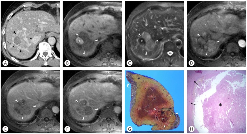
The dynamic contrast-enhanced CT images showed a 6 cm well-encapsulated mass in the right hepatic lobe, with the enhancement pattern of early arterial enhancement and delayed washout. And also, CT images showed enlargement of right and caudate lobe of the liver, and wall thickening and enhancement of both intrahepatic bile ducts (Fig. 1A). However, the biliary duct was not dilated. Based on these CT findings in addition to the increased level of CA19-9, the initial tentative diagnosis was a hepatic malignant tumor such as HCC or mass-forming cholangiocarcinoma with underlying alcoholic liver disease and intrahepatic cholangitis. Dynamic gadolinium-enhanced MR imaging was subsequently performed. On fat-suppressed T1-weighted image (Fig. 1B), the mass consisted of large area of hypointensity and small area of hyperintensity, which looked reversed with respect to signal intensity on fat-suppressed T2-weighted image (Fig. 1C). Small portion of T1 hyperintensity within the mass was interpreted to represent the presence of hemorrhage. The hypointense area of the mass on pre-contrast fat-suppressed T1-weighted image was hyperintense, slightly hypointense and hypointense, respectively, on gadolinium-enhanced T1-weighted arterial, portal, and delayed-phase MR images that were obtained at 30 seconds, 75 seconds and 180 seconds, respectively, after the injection of gadolinium (Figs. 1D-F). According to various imaging findings, such as early arterial enhancement and delayed washout, mosaic pattern of enhancement, internal hemorrhagic component, well defined tumoral capsule, and findings indicative of underlying alcoholic liver disease, the tumor was radiologically diagnosed as a HCC.
Subsequently, right hepatectomy was decided to perform as a curative treatment. In addition, conservative treatment such as administration of hepatotonic agents and antibiotics to treat underlying alcoholic liver disease and cholangitis had been done for 1 month before operation. The laboratory test performed just before the operation revealed that serum levels of total bilirubin (0.8 mg/dL), direct bilirubin (0.4 mg/dL), aspartate aminotransferase (51 IU/L), alanine aminotransferase (40 IU/L), gamma-glutamyl transpeptidase (101 IU/L), alkaline phosphatase (60 IU/L), and lactate dehydrogenase (441 IU/L) and CA19-9 (14.7 U/mL) were markedly improved. Right hepatectomy was performed at 6 weeks after admission, according to the initial treatment plan.
The gross surgical specimen showed that a 6 cm brown to reddish lobulated mass with both solid and cystic portions was surrounded by whitish fibrous capsule, and included hemorrhagic necrosis, necrotic debris and internal whitish septa (Fig. 1G). And also, multifocal irregular surface of the liver was demonstrated which was suggestive of chronic alcoholic liver disease. Microscopic examination revealed extensive areas of coagulative necrosis, infiltrations of chronic inflammatory cells including lymphoplasma cells and some eosinophils, fibrosis and focal areas of hemorrhage and hemosiderin pigments (Fig. 1H). These histologic features resulted in the diagnosis of hepatic abscess and chronic alcoholic liver disease. He had an uneventful postoperative course, and has been alive for 59 months after surgery.
The radiographic appearances of hepatic abscesses range from well circumscribed cystic lesion with enhancing rim to heterogeneously enhancing mass-like lesion, which is sometimes indistinguishable from hepatic neoplasm.2 However, in most cases, it is not difficult to diagnose hepatic abscess based on the imaging features by using US, CT or MRI.3 Further, even in cases of hepatic abscess mimicking malignant hepatic tumor, it could be straight-forward to correctly diagnose hepatic abscess with the aid of clinical symptoms and laboratory findings.
In our case, however, hepatic abscess was not preoperatively included in the differential diagnosis because imaging findings, such as early arterial enhancement and delayed washout, and mosaic pattern of enhancement, well-defined tumoral capsule with delayed enhancement, were consistently suggestive of HCC.4-6 In addition, there were no clinical symptoms or laboratory findings such as fever and leukocytosis that were suggestive of hepatic abscess. According to AASLD guideline,4 only CT or MR imaging can make a confirmative diagnosis of HCC without the need for histopathological confirmation. Thus, our case was preoperatively diagnosed to represent HCC according to typical imaging features at CT and MR imaging.
Hepatic abscess mimicking HCC has been rarely described in the literature.2,7 Brown et al2 reported cases of hepatic abscess mimicking HCC in the normal liver with contrast-enhanced CT, in which patients presented with high fever and leukocytosis. In those cases, hepatic abscesses were accompanied by portal vein thrombosis, and this finding favorably led to the diagnosis of abscess rather than HCC. Chou et al7 reported a case of hepatic abscess in the cirrhotic liver, which mimicked HCC at contrast-enhanced CT, presenting with high fever and marked elevated serum level of C-reactive protein. However, in contrast to previous reports, our case was very difficult to differentiate between hepatic abscess and HCC owing to a lack of clinical presentations suggestive of abscess.
In retrospect, in our case, follow-up imaging such as CT or MR imaging might be helpful to re-consider the initial diagnosis of HCC, and in turn, preoperative percutaneous ultrasound-guided biopsy should have been performed in a bid to avoid unnecessary surgery, given that liver cirrhosis was not clearly evident at both CT and MR imaging, and hepatic function was markedly improved at the laboratory test performed just before operation.
Hepatic abscesses are known to present with various imaging findings according to the degree of maturation and internal contents.1 Hepatic abscess rarely mimics hepatic neoplasm such as HCC. As evidenced by our case, if there are no clinical and laboratory findings suggestive of hepatic abscess, hepatic abscess that presents with imaging findings, such as early arterial enhancement and delayed washout, mosaic pattern of enhancement, and well-defined tumoral capsule with delayed enhancement could be hardly differentiated from HCC.
REFERENCES
1. Takayasu K, Okuda K. From Diagnosis to Treatment. New York: Oxford University Press; 1997. p. 299-307.
2. Brown KT, Gandhi RT, Covey AM, Brody LA, Getrajdman GI. Pylephlebitis and liver abscess mimicking hepatocellular carcinoma. Hepatobiliary Pancreat Dis Int 2003;2:221-225. 14599973.

3. Elizondo G, Weissleder R, Stark DD, Todd LE, Compton C, Wittenberg J, et al. Amebic liver abscess: Diagnosis and treatment evaluation with mr imaging. Radiology 1987;165:795-800. 2891154.


4. Bruix J, Sherman M. Management of hepatocellular carcinoma: An update. Hepatology 2011;53:1020-1022. 21374666.


5. Rimola J, Forner A, Tremosini S, Reig M, Vilana R, Bianchi L, et al. Non-invasive diagnosis of hepatocellular carcinoma Ōēż2 cm in cirrhosis. Diagnostic accuracy assessing fat, capsule and signal intensity at dynamic mri. J Hepatol 2012;56:1317-1323. 22314420.


Figure┬Ā1
Hepatic abscess mimicking HCC in a 70-year-old man with a long history of alcohol abuse. Axial contrast-enhanced portal phase CT image shows a 6 cm well-capsulated heterogeneously enhancing mass (arrowheads) with mosaic pattern of enhancement within the enlarged right hepatic lobe. CT image also demonstrates strong enhancement of thickened wall of intrahepatic ducts (arrows), suggestive of cholangitis (A). Axial T1-weighted fat-suppressed MR image shows that a 6 cm lobulated mass (arrowheads) in the right hepatic lobe, which consists of large area of hypointensity and small area of hyperintensity (asterisk) (B). Axial T2-weighted fat-suppressed MR image shows that a mass (arrowheads) in the right hepatic lobe consists of large area of hyperintensity and small area of hypointensity (asterisk), which looks reversed with respect to signal intensity on B (C). Axial gadolinium-enhanced T1-weighted arterial (D), portal (E), and delayed-phase (F) MR images show early arterial enhancement of the tumor that is accompanied by portal and delayed wash-out (arrowheads) with overally mosaic pattern of enhancement. And also, well defined tumoral capsule with delayed enhancement is seen on F (D-F). Photograph of gross specimen shows a 6 cm brown to reddish lobulated solid and cystic mass with surrounding whitish fibrous capsule (arrowheads), hemorrhagic necrosis (asterisk), necrotic debris and internal fibrous septa (arrows) within the resected right hepatic lobe. Note focal non-smooth surface (curved arrow) of the liver, which is suggestive of chronic alcoholic liver disease (G). Photomicrograph shows a necrotic lesion (asterisk) with surrounding fibrous capsule (arrows), which consists of extensive areas of coagulative necrosis, infiltrations of chronic inflammatory cells including lymphoplasma cells and some eosinophils, and fibrosis (H) (Hematoxylin and Eosin stain, ├Ś40).

- TOOLS
-
METRICS

- Related articles
-
MAFLD-related hepatocellular carcinoma in China: AnŌĆéincreasing problem


 PDF Links
 PubReader
 ePub Link
 Full text via DOI
 Full text via PMC
 Download Citation
 Print
Print


