Noninvasive serum biomarkers for liver fibrosis in NAFLD: current and future
Article information
Abstract
In the last 20 years, noninvasive serum biomarkers to identify liver fibrosis in patients with non-alcoholic fatty liver disease (NAFLD) have been developed, validated against liver biopsy (the gold standard for determining the presence of liver fibrosis) and made available for clinicians to use to identify ≥F3 liver fibrosis. The aim of this review is firstly to focus on the current use of widely available biomarkers and their performance for identifying ≥F3. Secondly, we discuss whether noninvasive biomarkers have a role in identifying F2, a stage of fibrosis that is now known to be a risk factor for cirrhosis and overall mortality. We also consider whether machine learning algorithms offer a better alternative for identifying individuals with ≥F2 fibrosis. Thirdly, we summarise the utility of noninvasive serum biomarkers for predicting liver related outcomes (e.g., ascites and hepatocellular carcinoma) and non-liver related outcomes (e.g., cardiovascular-related mortality and extra hepatic cancers). Finally, we examine whether serial measurement of biomarkers can be used to monitor liver disease, and whether the use of noninvasive biomarkers in drug trials for non-alcoholic steatohepatitis can accurately, compared to liver histology, monitor liver fibrosis progression/regression. We conclude by offering our perspective on the future of serum biomarkers for the detection and monitoring of liver fibrosis in NAFLD.
INTRODUCTION
The global prevalence of nonalcoholic fatty liver disease (NAFLD) has been rising steadily since 2006 [1] and NAFLD is estimated to affect a quarter of the world’s adult population [2]. NAFLD represents a spectrum of liver fat-associated conditions that begins with liver fat accumulation and progresses to steatohepatitis, liver fibrosis and cirrhosis. Within that spectrum of liver disease, it is patients with F3 [3] fibrosis and F4 [3] cirrhosis who are at substantial risk of death from end-stage liver disease and liver cancer. However, the earlier stages of liver fibrosis lend themselves well to therapeutic interventions to either attenuate or ameliorate progression and potentially reverse liver damage [4-7]. Thus, managing patients with NAFLD necessitates identification of F1 [3] and F2 [3] stages and estimation of the risk of progression to a more advanced stage of fibrosis/cirrhosis. However, liver disease can be hard to identify before it has reached a very advanced stage because it usually progresses without signs or symptoms [8].
In the last 20 years significant advances have been made in the development of noninvasive serum biomarkers for the identification of liver fibrosis. In this brief review, we describe these biomarkers and discuss their current utility and their potential future use in clinical practice. We consider whether liver fibrosis biomarkers have a role in: a) identifying F2 (that might be amenable to treatment as a relatively early stage of fibrosis), b) predicting patient outcomes and c), whether biomarkers can be used to help track progression or amelioration of liver fibrosis.
INITIAL AND CURRENT USE OF NONINVASIVE SERUM BIOMARKERS FOR NAFLD
Liver fibrosis is one of the most relevant prognostic factors for important clinical outcomes in NAFLD [9], yet liver fibrosis often remains undiagnosed until it has progressed to cirrhosis. With the global prevalence of NAFLD estimated to be between 31.6% and 40.8% of the population [10], it is important to be able to detect liver fibrosis early in the disease process, so that effective interventions can be implemented before the disease becomes too advanced. The gold standard for identification and staging of liver fibrosis is liver biopsy, however, it is a diagnostic procedure that is time consuming, costly, invasive, subject to sampling error [11], and not scalable considering the magnitude of the global health care burden imposed by NAFLD.
Noninvasive serum biomarkers for fibrosis were initially developed by and for secondary care physicians, to use as a diagnostic assessment tool to detect patients who have advanced liver fibrosis and/or cirrhosis, offering an alternative and potential replacement to liver biopsy. A number of noninvasive serum biomarkers have been developed over the last 20 years and we now have tests, that have been validated against liver biopsy, such as the enhanced liver fibrosis (ELFTM) test [12], fibrosis-4 (FIB-4) index [13], NAFLD fibrosis score (NFS) [14], aspartate aminotransferase to platelet radio index (APRI) [15] and FibroTest® [16] (FibroSURETM in the USA). These relatively common tests are widely available for use in both primary and secondary care and offer a variable degree of accuracy and reliability (Table 1).

Summary of the performance comparison of five widely available and frequently used noninvasive serum biomarkers for diagnosing ≥F3 liver fibrosis in NAFLD
Combining noninvasive serum biomarkers has been shown to further improve diagnostic performance compared with single biomarker performance alone [17,18]. Nevertheless, the current use of noninvasive serum biomarkers focuses on excluding disease, e.g., stratification of patients into those who have a high probability of ≥F3 fibrosis versus those who have a low probability of ≥F3 fibrosis. The utility of noninvasive serum biomarkers is therefore limited because even though they have been used to identify someone with a high probability of ≥F3 fibrosis, additional tests are required to confirm this. For example, in UK primary care, the biomarkers NFS, FIB-4 and ELFTM are recommended for use to identify patients with a high probability of ≥F3 fibrosis [19] but as the biomarker itself is not informative enough as a basis for intervention, the recommendation is to follow biomarker testing with vibration controlled transient elastography (VCTE) [20], to confirm the stage of fibrosis. In Korea, the recommendation is to assess for fibrosis using radiological examinations such as VCTE [21]. If this is not feasible then NFS or FIB-4 are the recommended tests [21].
DO BIOMARKERS HAVE A ROLE IN IDENTIFYING F2 FIBROSIS?
We now know that F2 fibrosis has important consequences for patients [22,23]. F2 fibrosis is a risk factor for cirrhosis and overall mortality and F2 increases the risk of extra hepatic complications including cardio vascular disease [22,23]. Approximately 20% of patients diagnosed with low-levels of liver fibrosis (F1–F2) will progress to F3, or F4, within 5 years [24]. F2 is a stage of fibrosis that is easily managed in primary care and it is potentially treatable and maybe halted or reversed through lifestyle changes [6,25,26]. Alternatively, medications such as anti-fibrotic therapeutic drugs (currently in phase 3 trials [27]) or glucagon-like peptide-1 agonist medication [28] may have beneficial effects on the early stages of liver fibrosis. It is therefore important for clinicians to be able to identify F2 accurately, precisely, quickly and easily, which noninvasive serum biomarkers have the potential to do. However, there are difficulties in determining the optimum cut-off value to use to differentiate intermediate states of fibrosis from the more advanced stages [29,30]. To date no one biomarker is recommended for the detection of F2 [13,31].
Recent systematic reviews evaluating the five widely available noninvasive biomarkers concluded that APRI [32], FIB-4 [32], FibroTest® [33] and NFS [32] showed a fair [34] performance for identifying ≥F2 fibrosis (Table 2). The performance of ELFTM [35] however was evaluated as good [34], although it should be noted that ELFTM may produce a high number of false positive tests (specificity=12%). In another systematic review, PRO-C3 [36] (N-terminal type III collagen pro-peptide) a less widely available noninvasive blood biomarker, has been shown to match the performance of ELFTM and outperform APRI, FIB-4, FibroTest®, and NFS [32]. In this study PRO-C3 had a sensitivity and specificity of 68% (95% confidence interval [CI], 0.50–0.82) and 79% (95% CI, 0.71–0.86) respectively, with an area under the curve (AUC) of 0.81 (95% CI, 0.77–0.84) [36]. However, the availability of PRO-C3 is limited. Currently, the PRO-C3 assay is exclusively produced by a pharmaceutical company and at present is only used for research purposes and is not recommended for clinical use [36].

Comparison of the performance of ELF™, FIB-4, APRI, FibroTest®, and NFS for identifying ≥F2 fibrosis
Ideally, clinicians should be able to quickly and easily assess their patients for ≥F2 fibrosis without having to request additional costly blood tests that require specialist evaluation (e.g., ELFTM and FibroTest®). Sripongpun et al. [37] developed and validated a biomarker (Steatosis-Associated Fibrosis Estimator, SAFE) specifically to identify ≥F2 fibrosis. SAFE has seven variables (sex, body mass index [BMI], diabetes status, aspartate transaminase [AST], alanine transaminase [ALT], platelet and globulin) [37]. SAFE is therefore similar to the NFS that includes age, BMI, platelet count, AST and ALT ratio [14]. SAFE was shown to outperform NFS [37], suggesting that the coefficients applied to SAFE maybe a better fit for identifying ≥F2 fibrosis in modern NAFLD patients [37].
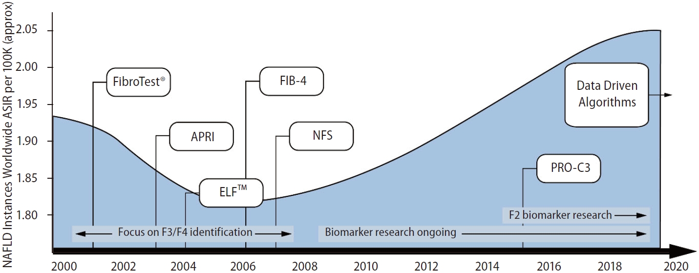
The use of machine learning from serum biomarker data has been found to offer a good performance for identifying ≥F2 fibrosis, AUC 0.86 [38]. A recently published study utilised routinely available data to develop and validate six algorithms (LiverAID XXS, XS, S, M, L, and 4XL) to identify ≥F2 [38]. The diagnostic performance of all the LiverAID models for detecting ≥F2 outperformed FIB-4 and APRI, and in all cases was statistically significant (P≤0.001): the AUC of LiverAID-XXS=0.86, the AUC of LiverAID-XS=0.89, the AUC of LiverAID-S=0.91, the AUC of LiverAID-M=0.92, the AUC of LiverAID-L=0.92, the AUC of LiverAID-4XL=0.94, the AUC of FIB-4=0.70 and the AUC of APRI=0.74. This demonstrates how machine learning models can utilise data and very quickly learn to identify liver fibrosis. However, the performance of machine learning algorithms is dependent on the quantity and quality of the input data and using liver biopsy as the reference standard. To date, the data available from liver histology studies are not sufficient to develop and guide the algorithms and available datasets are currently far too small [39]. At present, the use of machine learning to identify fibrosis is still in its infancy. That said, machine learning is well positioned to deal with this type of dynamic data in the future (Fig. 1) [40].

Timeline showing the global rise in NAFLD and the emergence of noninvasive biomarkers for fibrosis in NAFLD. NAFLD, non-alcoholic fatty liver disease; ASIR, age-standardised incidence rate per 100,000 persons; ELFTM, enhanced liver fibrosis; FIB-4, fibrosis-4; NFS, nonalcoholic fatty liver disease fibrosis score; APRI, aspartate transaminase to platelet ratio index; PRO-C3, type III collagen marker of the N-terminal pro-peptide.
CAN A SINGLE BIOMARKER TEST PREDICT PATIENT OUTCOMES?
Observational studies have shown biopsy-confirmed liver fibrosis is a prognostic factor for patients with NAFLD [41,42]. A single biomarker that can predict patient outcomes as well as, or better, than liver biopsy would be a useful tool for clinicians managing patients with liver disease. However, there is conflicting evidence [43-45] and this may be in part due to the ethnicity of populations studied, the length of follow-up period, or inadequate sample sizes and the limited power of the studies to address these questions [43-45].
A medium sized study (n=153) based in Israel [43], with a follow-up period of 100 months, has shown that FIB-4 and NFS, but not APRI, when compared with liver biopsy, are good predictors of overall mortality. Higher FIB-4, NFS and APRI scores were also associated with hepatic and extra-hepatic malignancies [43]. A larger sized study (n=301) in Japan with a follow-up period of 84 months, has shown that FIB-4 and NFS are useful for predicting the occurrence of liver-related complications (e.g., varices, ascites or encephalopathy) [44]. However, these scores were limited in their ability to predict extrahepatic malignancies [44]. A recent systematic review concluded that in secondary care, FIB-4, NFS and APRI show limited performance in predicting changes in fibrosis (as evaluated by biopsy) [45]. However, these scores consistently predicted liver-related morbidity (e.g., ascites, esophageal varices or hepatocellular carcinoma), and also liver-related mortality [45].
A more recent (2022) systematic review and meta-analysis has reaffirmed that NFS and FIB-4 are reliable and comparable to liver biopsy as prognostic markers of all-cause mortality in NAFLD patients. Additionally, NFS may be useful for predicting risk of cardiovascular death [46]. Further, a large retrospective study (n=5,123) in America [47] found that the risk of progression to cirrhosis and decompensation increased by FIB-4 strata at NAFLD diagnosis [47]. In Individuals with FIB-4 <1.3, the risk of NAFLD progression was higher than for those with 1.30-2.67 (hazard ratio [HR]=3.67; 95% CI=1.65–8.15; P=0.0014) and FIB-4 >2.67 (HR=56.26; 95% CI=25.77–122.83; P<0.001) [47]. Also, the risk of death was higher in individuals with FIB-4 >2.67 (HR, 3.26; P<0.001) [47]. In a different study, it has been shown that ELFTM predicts clinical outcomes more accurately than liver biopsy [48]. A one-point increase in ELFTM score was associated with a twofold increase in risk of liver-related clinical outcome (defined as liver-related death or episode of decompensated cirrhosis e.g., ascites or esophageal variceal hemorrhage) [48]. Therefore, noninvasive serum biomarkers for liver fibrosis in NAFLD, e.g., NFS, FIB-4, and ELFTM may help predict non-liver-related outcomes e.g., cardiovascular-related mortality [46], and extra-hepatic cancers [43,44]; thus demonstrating their utility beyond simply diagnosing liver disease.
In the US, ELFTM has been granted marketing authorization by the American Food and Drug Administration (FDA) for use as a prognostic risk assessment tool for assessing the likelihood of fibrosis progression in patients with advanced fibrosis [49]. The guidance from the manufacturers of ELFTM is that in patients with F3 bridging fibrosis, an ELFTM score of ≥9.8 indicates an increased risk of progression to cirrhosis in 1–5 years [50]. The guidance also states that in patients with compensated cirrhosis, an ELFTM score of ≥9.8 indicates an increased risk of progression within 5 years to a liver-related event (e.g., development of hepatocellular carcinoma, liver failure or death) [50]. The manufacturers of ELFTM do not, however, quantify how great the risk of progression is. In our opinion, a more accurate interpretation of their guidance should be that after a liver biopsy has diagnosed F3 bridging fibrosis, an ELFTM score of ≥9.8 indicates a risk of progression to cirrhosis in 1–5 years. In the UK, the ELFTM test is the recommended noninvasive blood biomarker test, to identify advanced fibrosis in patients diagnosed with NAFLD [20]. The guidelines are to repeat ELFTM every three years [20], and not to use serial ELFTM measurements to monitor disease progression. Rather, the test should be used at any single moment in time to predict risk of prevalent ≥F3 liver fibrosis.
CAN SERIAL MEASUREMENT OF LIVER FIBROSIS BIOMARKERS HELP TRACK OR MONITOR DISEASE PROGRESSION?
As it is often uncertain how quickly liver disease will progress, a reliable noninvasive test to monitor progression over time is needed. Noninvasive serum biomarkers have the potential to monitor disease progression or amelioration over time. Having a baseline biomarker result that is repeated at regular intervals to monitor liver health would be useful for both patients and clinicians. However, repeating a biomarker and relying on the result to inform a prognosis requires the change in biomarker score to be independently validated against the change in liver biopsy, the gold standard for determining the presence and degree of liver fibrosis.
An alternative to using liver biopsy to validate biomarker score changes would be to examine retrospective biomarker scores over time in relation to liver disease progression, as was undertaken by Hagström et al. [51]. These investigators used data from a retrospective population based cohort (1986–1996) and showed that repeating FIB-4 within a 5-year period can, in comparison to a single measurement, help identify individuals who are at a higher risk of developing severe liver disease [51]. These authors noted that repeating FIB-4 is only recommended for individuals at a low risk of worsening fibrosis. The recommendation for a high risk patients was that these individuals should undergo additional diagnostic testing, e.g., VCTE, without repeat testing of FIB-4 [51]. In another retrospective analysis, Balkhed et al. [52] examined data from a high prevalence of liver disease setting and showed the accuracy of FIB-4 (and APRI) is only weakly associated with disease progression. The authors concluded that the biomarkers have limited clinical utility in monitoring the course of NAFLD progression [52].
Metabolomics analysis has been used as a promising method in NAFLD to investigate novel biomarkers involved in the pathogenesis of the disease [53]. In particular, serum lipocalin 2 has been identified as a key molecule participating in transport of fatty acids [54], that may serve as a valuable NAFLD biomarker for monitoring the initiation and progression of fibrosis [54].
Currently, there is still no licensed drug treatment for NAFLD. In the last decade, there have been many clinical trials testing new drugs for the treatment of liver disease in NAFLD. However, data obtained from these trials have shown suboptimal results, particularly for treatment of liver fibrosis [55]. In clinical trials for NAFLD treatment, liver biopsy is the reference standard used to assess liver fibrosis, which means that participants are required to have at least two (baseline and end of study) invasive procedures to assess the efficacy of a drug. In therapeutic drug trials for non-alcoholic steatohepatitis (NASH), noninvasive serum biomarkers are often (but not always) included to assess for changes in liver fibrosis. Therefore, when the liver biopsy findings in a drug trial show a change in the staging of fibrosis, the performance of biomarkers can be compared against the changes in liver histology.
We reviewed all 21 of the NASH drug trials from a recent systematic review and meta-analysis by Ampuero et al. [55] (Supplementary Table 1). Five [27,56-59] studies did not use any widely available noninvasive biomarker to assess changes in liver fibrosis, one [60] study stated that the data is not publicly available, and two [61,62] were conference reports/poster presentations. We tabulated the remaining 13 studies [63-75], (Supplementary Table 2) and an abridged version shown as Table 3, to illustrate the biopsy-observed changes in liver fibrosis and the changes that occurred in serum biomarker scores (ELFTM, NFS, APRI, FIB-4, FibroTest® , and PRO-C3) between baseline and follow-up assessment. It should be noted that the primary aim of the drug trials shown in the tables was to evaluate the efficacy of a therapeutic drug treatment for NASH, rather than to investigate the ability of noninvasive serum biomarkers to monitor change in histological measurement of fibrosis. As such, the value of the data reported and available from the published research papers is limited to address the question of whether biomarkers can be used to monitor changes in fibrosis attributed to a therapeutic intervention. For example, the biomarker scores at baseline and follow-up for ELFTM, NFS, APRI, FIB-4, FibroTest®, and PRO-C3 in all the trials were all reported as an average score observed changes between baseline and follow up. Nine [63-71] of the studies included participants with F1 and F2 (and in some studies F0); yet the serum biomarkers used to assess fibrosis (ELFTM, NFS, APRI, FIB-4, and FibroTest®) are currently only validated for ≥F3 fibrosis. The participant eligibility criteria for the remaining four [72-75] studies was F3 at baseline. Therefore a comparison of biomarker performance against changes in liver histology should be possible. However, only one of the studies (Harrison et al. [74], 2020) provided sufficient data to make this comparison. Therefore, the utility of noninvasive biomarkers to track changes in liver fibrosis needs further study in therapeutic trials targeting treatment of fibrosis.
CONCLUSION
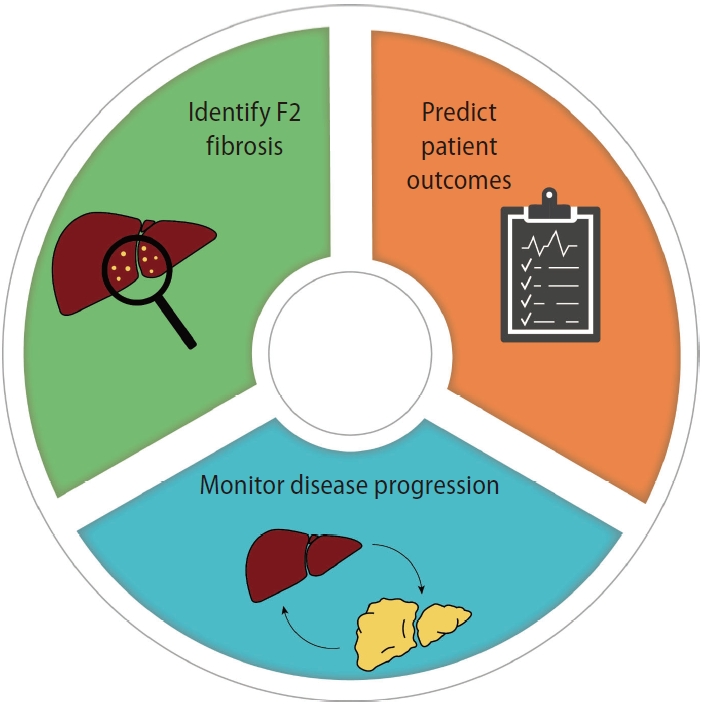
The current use of widely available noninvasive serum biomarkers for fibrosis in NAFLD continues to be used to identify patients who have a high probability of ≥F3 fibrosis in settings where there is a high prevalence of more severe liver disease. It remains uncertain whether biomarkers have sufficient sensitivity and specificity to be able to monitor progression in fibrosis, or amelioration of fibrosis with therapeutic interventions. Although there is a recognized need to identify fibrosis earlier in the disease process, no single biomarker has been shown to be accurate or precise enough to identify patients with F2 liver fibrosis. Increased liver fibrosis biomarker scores are associated with liver-related morbidity and mortality and also associated with an increased risk of non-liver related patient outcomes. Currently, there is an insufficient evidence to demonstrate that a change in a biomarker score allows prediction of a change in liver fibrosis. Finally, we consider that it is now crucial to develop biomarkers that accurately and precisely identify F2, and to continue to investigate whether biomarkers can be used for assessing and monitoring disease progression/regression with therapeutic interventions that include both drugs and lifestyle change (Fig. 2).
Notes
Authors’ contribution
All authors (Tina Reinson, Ryan M. Buchanan, and Christopher D. Byrne) contributed to the review structure and concept; drafting of the manuscript and its critical revision; and approved the final version.
Conflicts of Interest
The authors have no conflicts to disclose.
Acknowledgements
For the purpose of Open Access, the author has applied a Creative Commons Attribution (CC BY) licence to any Author Accepted Manuscript version arising from this submission. The authors would like to thank the NIHR Southampton Biomedical Research Centre and the University of Southampton for their support.
CDB and RMB are supported in part by the Southampton NIHR Biomedical Research Centre (IS-BRC-20004), UK.
SUPPLEMENTARY MATERIAL
Supplementary material is available at Clinical and Molecular Hepatology website http://www.e-cmh.org).
Brief summary of the 21 therapeutic drug trials examined in Ampuero et al.’s 2022 meta-analysis of variables influencing the interpretation of clinical trial results in NAFLD[22]
Comparison between change in noninvasive serum biomarkers and change in liver fibrosis assessed by liver histology, in therapeutic trials of nonalcoholic steatohepatitis (NASH)
Abbreviations
APRI
aspartate transaminase to platelet ratio index
AUC
area under the curve
CI
confidence interval
CVD
cardio vascular disease
ELF™
enhanced liver fibrosis test
FDA
Food and Drug Administration
FIB-4
fibrosis-4 index
GLP-1
glucagon-like peptide-1
METAVIR
meta-analysis of histological data in viral hepatitis
NAFLD
nonalcoholic fatty liver disease
NASH
nonalcoholic steohepatitis
NFS
NAFLD fibrosis score
NPV
negative predictive value
PPV
positive predictive value
PRO-C3
type III collagen marker of the N-terminal pro-peptide
VCTE
vibration controlled transient elastography


